References

Qazi SH, Khurram MS, Hassan M Orthodontic record keeping: a study. Pakistan Oral Dent J. 2010; 30:432-435
Arshad F, Narendranath V, Sarala KS Knowledge and practice of orthodontic record keeping - a clinical audit. Ann Int Med Dent Res. 2022; 8:45-56 https://doi.org/10.53339/aimdr.2022.8.5.7

A simple way to take the best mid-treatment orthodontic model

From Volume 17, Issue 4, October 2024 | Page 179

Authors

Anusha Hegde

Department of Orthodontics and Dentofacial Orthopedics, SDM College of Dental Sciences and Hospital, A constituent unit of Shri Dharmasthala Manjunatheswara University, Sattur, Dharwad, Karnataka, India.

Articles by Anusha Hegde

Email Anusha Hegde

Yasodipta Subhadarshini

Department of Orthodontics and Dentofacial Orthopedics, SDM College of Dental Sciences and Hospital, A constituent unit of Shri Dharmasthala Manjunatheswara University, Sattur, Dharwad, Karnataka, India.

Articles by Yasodipta Subhadarshini

Email Yasodipta Subhadarshini

Apurba Gupta

Department of Orthodontics and Dentofacial Orthopedics, SDM College of Dental Sciences and Hospital, A constituent unit of Shri Dharmasthala Manjunatheswara University, Sattur, Dharwad, Karnataka, India.

Articles by Apurba Gupta

Roopak D Naik

BDS, MDS

Professor and Head, Department of Orthodontics and Dentofacial Orthopaedics, SDM College of Dental Sciences and Hospital, Sattur, Dharwad – 580 009 Karnataka, India

Articles by Roopak D Naik

Article

Diagnostic records are very important in orthodontics. From diagnosis to treatment, planning is executed using proper records, including photographs, models and radiographs. Orthodontic study models have been the ‘gold standard’ with numerous benefits.1 Records find their application in patients’ education, self-appraisals, medicolegal issues and publications.2 The orthodontic model before bonding and after debonding are quite easy to obtain as no brackets are present. On the other hand, taking the mid-treatment model is another issue entirely. One sees tearing of impressions, air bubble formations, missing anatomical landmarks and many more.

Here is a simple method to avoid all these problems and get the best mid-treatment model. The steps are:
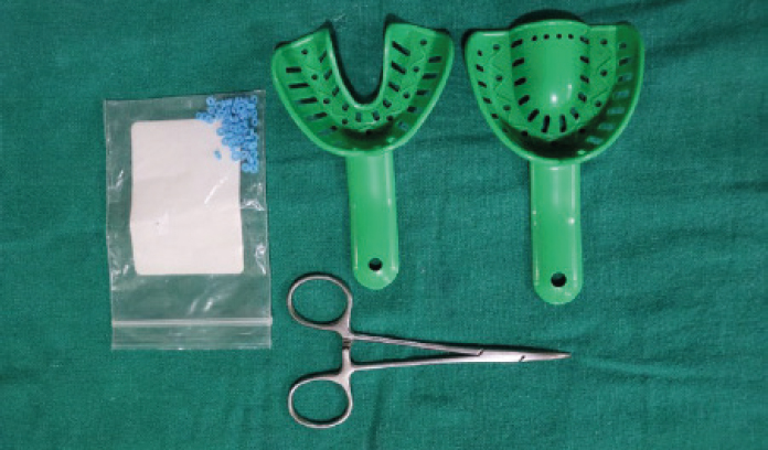
  • Armamentarium (Figure 1);
  • Remove the O-rings, E-chains or any other active component and orthodontic wire;
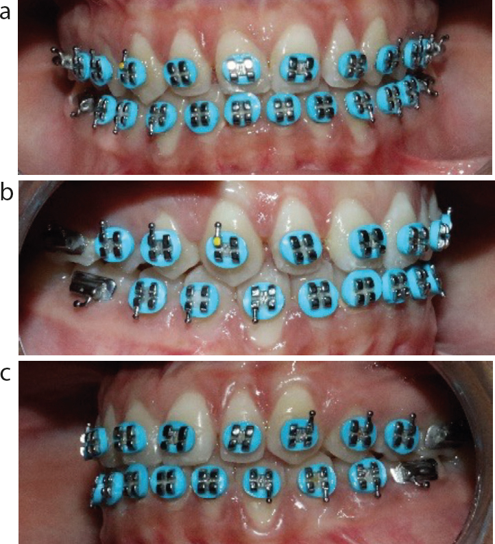
  • Place the separators (light blue) onto the bracket wings (Figure 2);
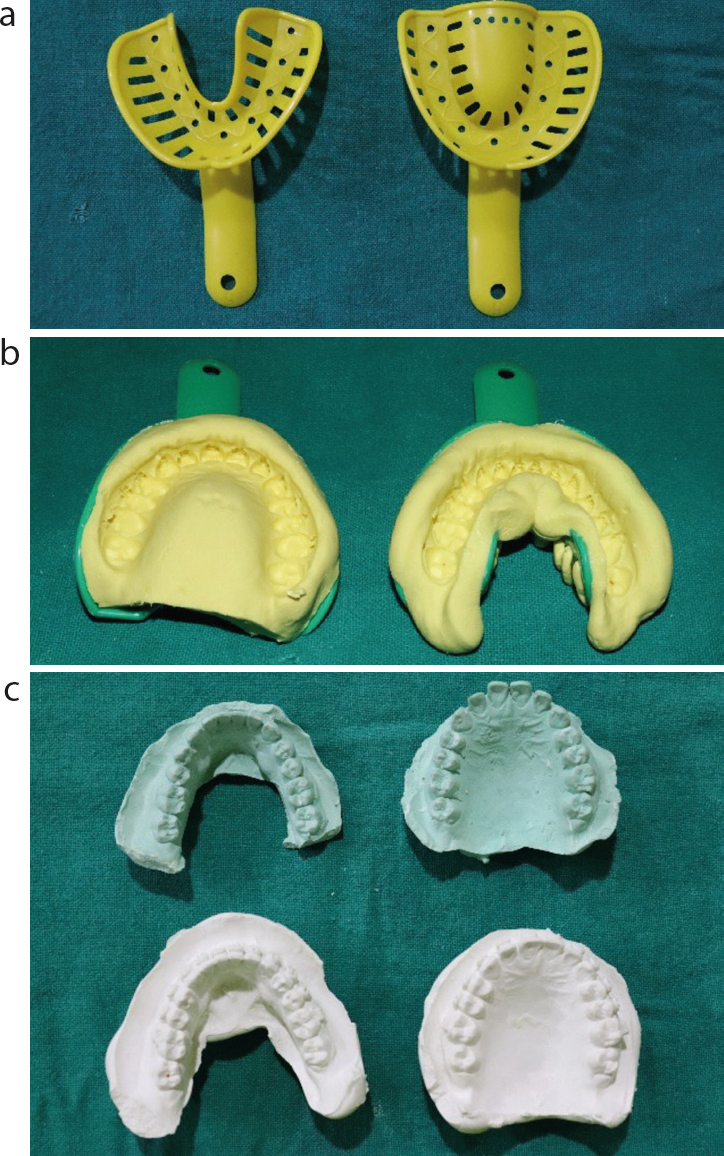
  • Take the alginate impression and pour using white type III dental gypsum (Figure 3);
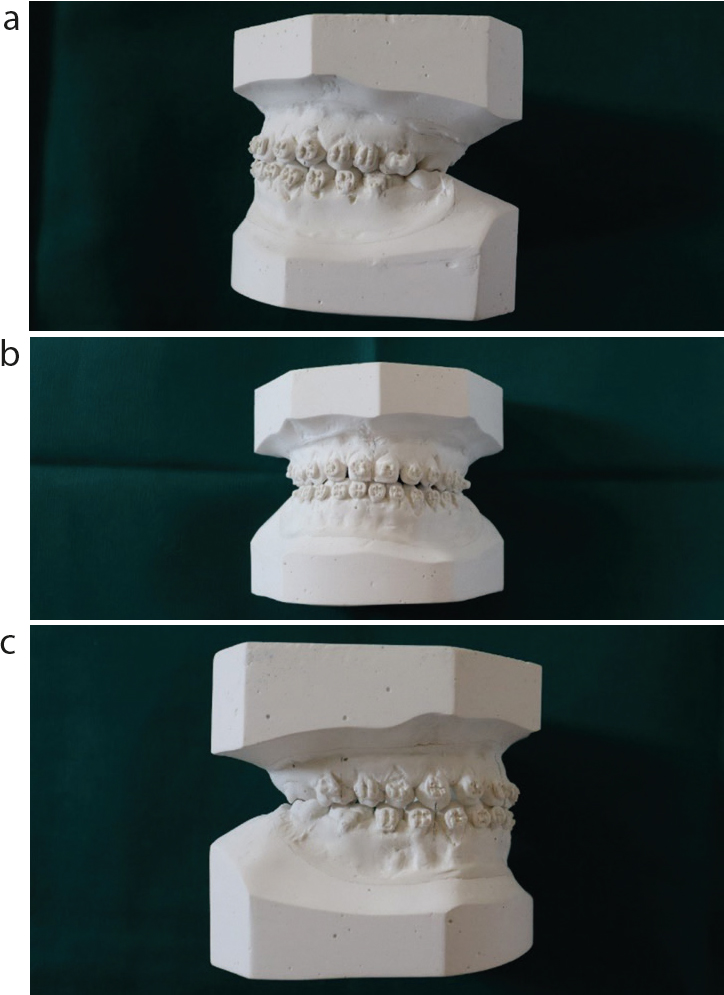
  • The perfect mid-treatment model is ready (Figure 4).
  • Figure 1. Armamentarium.
    Figure 2. (a–c) Blue separators placed onto the bracket wings.
    Figure 3. (a–c) Impressions.
    Figure 4. (a–c) Completed mid-treatment model.

    Advantages of this method:

  • Easy availability of material;
  • Chairside method;
  • Easy clinical procedure;
  • Less time is needed;
  • Cost-effective;
  • Better compliance from patients;
  • Perfect mid-treatment model procured.