Abstract
Ectopic maxillary canines are present in 1–3% of the population and may consequentially cause root resorption in 66.7% of lateral incisors. Ectopic canines are thought to be of polygenic and multifactorial origin, although
From Volume 14, Issue 4, October 2021 | Pages 195-199
Ectopic maxillary canines are present in 1–3% of the population and may consequentially cause root resorption in 66.7% of lateral incisors. Ectopic canines are thought to be of polygenic and multifactorial origin, although

Ectopic maxillary permanent canines occur in 1–3% of the population, over two-thirds of which are impacted palatally.1–3 Despite the frequency of ectopic canines, they are often missed in routine dental examinations and, as a result, are often not referred until permanent and irreversible resorption has occurred. Although there is no single cause known, two common theories for maxillary canine impaction include the guidance theory and the genetic theory.4 A polygenic, multifactorial genetic pattern of inheritance of palatally displaced canines was suggested by Peck and Peck.5 Similarly, hypodontia is thought to have a multifactorial origin and incidence varies from 2.6% to 11.3% of the population.6 After the wisdom teeth, it most frequently affects the second premolars and lateral incisors. Interestingly, the relationship between ectopic canines and hypodontia has been investigated in several studies, particularly detailing the relationship between ectopic canines and agenesis of lateral incisors based on the guidance theory. There are fewer studies that detail a relationship between palatally ectopic canines and agenesis of the second premolars.7–9 Furthermore, resorption of incisors adjacent to ectopic canines may be commonly expected sequelae. This report demonstrates the similar presentation of such resorption, but also idiopathic resorption of the lateral incisors on the contralateral side. A genetic link for internal and external root resorption has been suggested in the literature to be the result of genetic polymorphism of the interleukin (IL-1) mutation.10,11 This report of monozygotic twin boys who attended the orthodontic department with palatally ectopic canines, agenesis of one of more second premolars and root resorption of both adjacent and contralateral teeth may, therefore, add to the evidence suggesting a genetic susceptibility for root resorption.
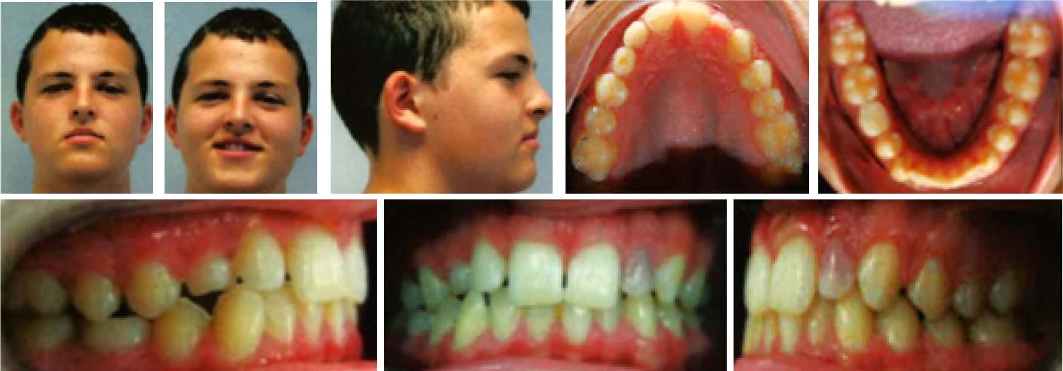
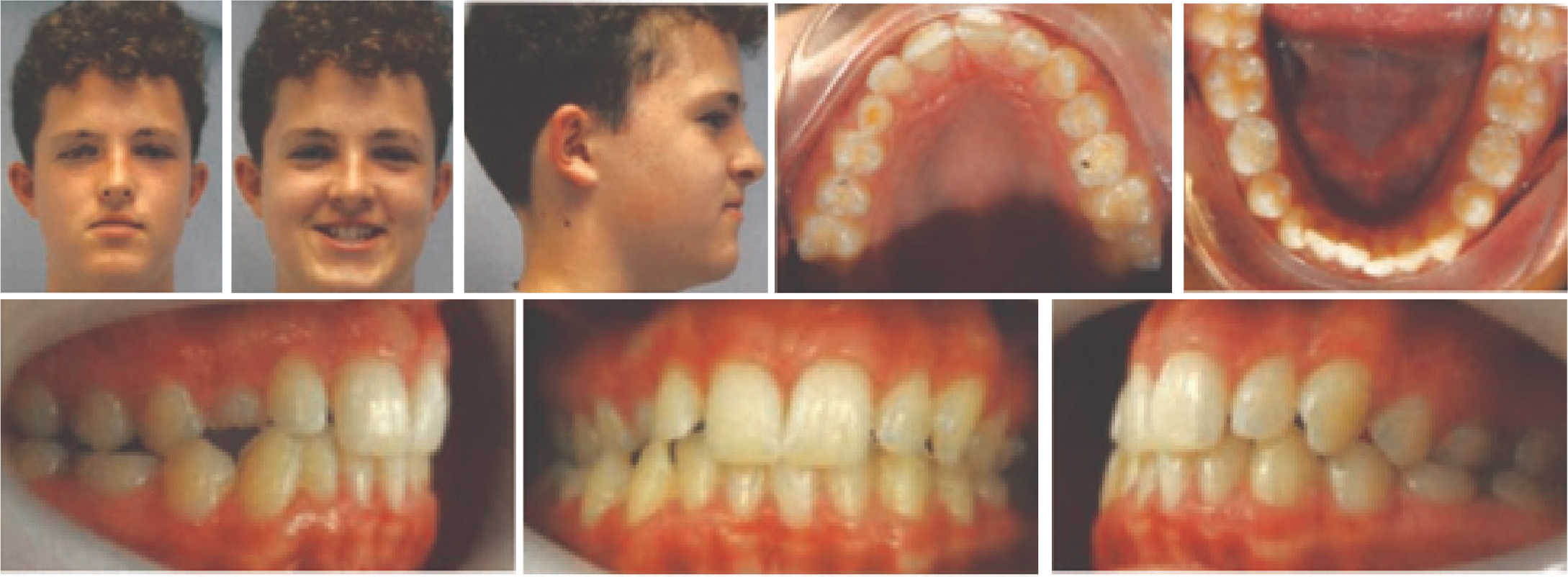
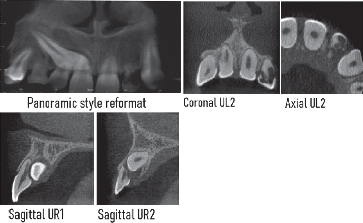
At age 14 years, Twin 1 was referred to the orthodontic department by his general dental practitioner (GDP) for the impacted UR3, retained LRE and non-vital UL2. Medically, he was fit and well, although he had an allergy to amoxicillin, was born prematurely at 34 weeks and had bronchiolitis as a baby. His chief complaint was the discolouration of UL2.
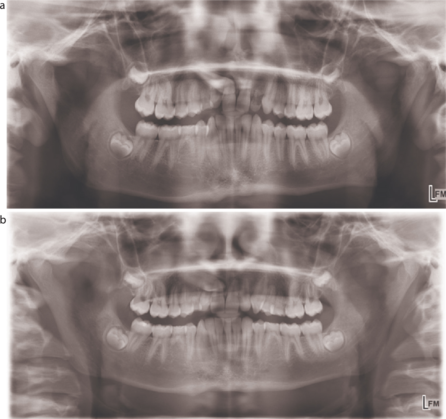
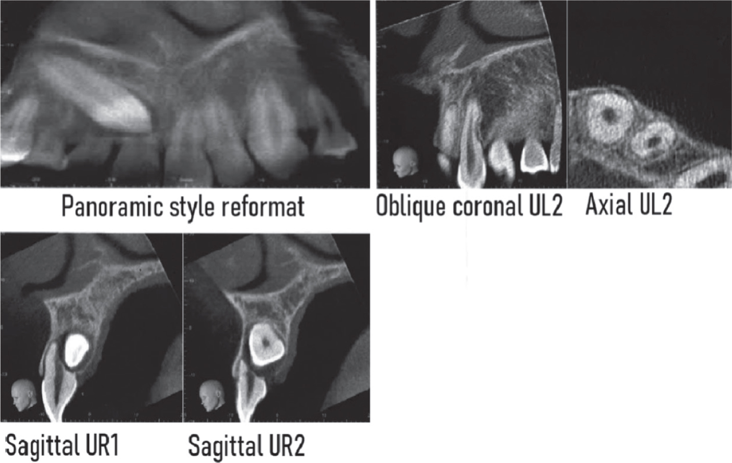
Twin 2 was also referred to the orthodontic department at age 14 years by his GDP for the impacted UR3 and multiple retained retained second primary molars. He had no complaints and was medically fit and well, although he had been born prematurely at 34 weeks and also had bronchiolitis as a baby. Table 1 outlines the assessment of the twins.
| Twin 1 | Twin 2 | |
|---|---|---|
|
Extra-oral assessment
|
Mild Class 3 skeletal base |
Mild Class 3 skeletal base |
|
Intra-oral assessment
|
Palatally ectopic and impacted UR3 |
Palatally ectopic and impacted UR3 |
| Class 1 incisal relationship |
Class 1 incisal relationship |
|
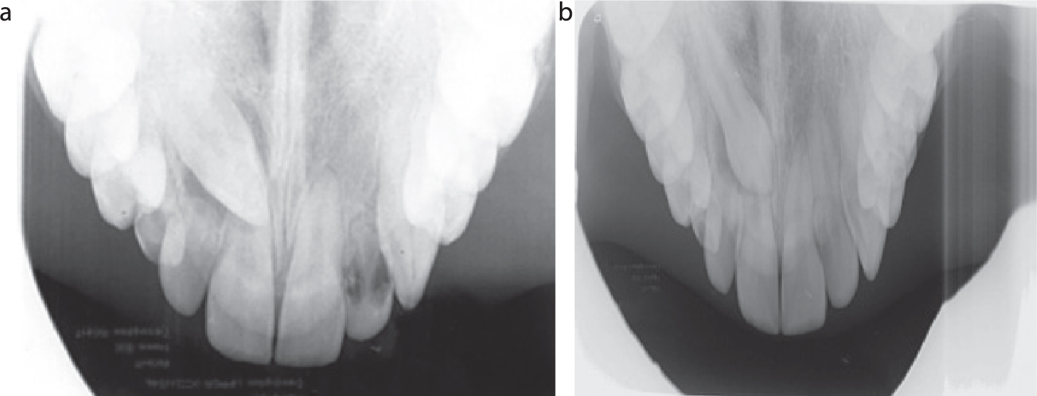
| Radiographic assesment | Upper standard occlusal (USO) (Figure 3) |
Upper standard occlusal (USO) (Figure 3) |
| Lateral cephalogram |
Lateral cephalogram |
|
| CBCT (Figure 5) |
CBCT (Figure 6) |
Twin studies have historically been a useful means of assessing the impact of nature versus nurture.12 Many case reports describe monozygotic twins presenting with hypodontia or ectopic canines; however, this report uniquely demonstrates both anomalies, with the additional feature of root resorption in this pair of genetically identical twins. Furthermore, hypodontia of the premolars rather than lateral incisors contrasts to the guidance theory and strongly suggests a genetic aetiology of these anomalies. Similar observations were seen in a case report by Leonardi et al describing palatally displaced canines in twin females.13
The homeobox genes are thought to be responsible for the development of teeth, including Msx1, Msx2, Dlx1, Dlx2, Shh and Pax-9. Msx1 and Msx2 are responsible for the developmental position and further development of tooth buds. Mutation in genes and disruption of regulatory molecules may result in anomalies in the dentition.14 Arte et al assessed 214 family members and found that incisor–premolar agenesis is inherited as an autosomal dominant trait, and is responsible for anomalies including ectopic canines.9 Although further research with larger cohorts is needed to establish the relationships further, it does reinforce the importance of early referral and screening of identical twins for similar pathology.
Owing to incomplete penetrance, however, a genetic association may not always be established at a routine dental examination, emphasizing the need for a thorough assessment. It is important to recognize and seek early referral for patients with hypodontia, especially when the primary teeth are retained, to prevent infra-occlusion and establish long-term prognosis and future restorative plans.
Patients between the ages of 10 and 11 years should be screened for possible ectopic or missing canines in the mixed dentition by initially palpating buccally.15 When evaluating a patient, a practitioner should consider several factors, including the amount of space available for the canine, the morphology and position of adjacent teeth, bone contour, mobility of primary or permanent teeth and radiographic appearance.
With the increased availability of cone beam CT, it has become evident that up to 67% of lateral incisors may be damaged by ectopic canines.16 A further interesting, although unfortunate, aspect of this case includes the severity of cervical resorption affecting UL2 in Twin 1, despite the full eruption of the canine. Given the extent of the resorption on both the adjacent and contralateral incisors, consideration of a genetic susceptibility to resorption is emphasized in this case report and may be related to the presence of polymorphisms of IL-1, including expression of the IL-1β plus C3953, and IL-1RN plus 2018C alleles.10,11 While most of the literature is based on susceptibility to external apical root resorption following orthodontics, this report suggests that the genetic susceptibility to root resorption may be present idiopathically. While the extent of the resorption may have a genetic element, the cause of the resorption was likely to be related to the path of eruption of the canine, which further emphasizes that early intervention is key in order to prevent catastrophic resorption and eventual loss of adjacent incisors. In addition, there is some evidence to suggest that early removal of the primary canines may, in fact, encourage the permanent canine to align, although this is controversial.17,18
There could be a genetic basis for root resorption seen in these identical twins and this subject deserves further investigation. It is important to recognize ectopia at an early age to minimize potential damage to the adjacent incisors and, where possible, prevent or reduce the patient's burden and cost of long-term fixed prostheses.
That referrals continue to be delayed highlights the need for further training of dentists on this topic.
