References
The aberrant canine part 1: aetiology and diagnosis
From Volume 10, Issue 4, October 2017 | Pages 126-130
Article

The permanent canine usually erupts uneventfully, but occasionally it may fail to do so. When this occurs there is a potential for the adjacent teeth to be damaged. Even when it does not cause any damage, treatment of the ectopically positioned canine can present a substantial challenge to the orthodontist. This paper presents a summary of the development and eruption of the permanent canine, both upper and lower, possible adverse effects of an aberrant position and the different treatment options.
Development of the permanent canine
Calcification of the upper and lower permanent canine teeth begins at 4 to 5 months post-partum, with crown formation being complete by the age of 5 years. The lower permanent canine erupts at around 10 years of age (± 6 months) and the upper canine at about 11.5 years (± 6 months). Although the upper permanent canine has a long path of eruption, the crown should be palpable beneath the mucosa in the buccal sulcus by the age of 10 years. Eruption is guided by the distal surface of the lateral incisor root in the case of the maxillary canine, and this can lead to the distal angulation of the lateral incisor. As a result, it is normal to see physiological spacing of the upper incisors in the mixed dentition (Figure 1), often referred to as the ‘ugly duckling’ stage, which then closes as the upper canines erupt, guided by the distal surfaces of the upper lateral incisor roots. Once the canines have fully erupted, the intercanine width in both the upper and lower arches is at its greatest and will then only reduce over time.1

Aetiology of the aberrant canine
Aberrant permanent canine position has been reported with an incidence of between 2%1and 5%,2 with over 90% of these teeth being maxillary canines.3 The majority of maxillary permanent canine impactions are either palatal (60−85%), or within the line of the arch.4 Initially, it was thought that persistence of the deciduous canine was the causative factor,5 but this is more likely to be a consequence of the ectopic crown position of the permanent tooth rather than the principal aetiological factor. There are two main theories to explain impaction of maxillary permanent canines, namely:
1. The Guidance Theory.6 It is thought that the crown of the upper canine is guided into position within the arch by the root of the lateral incisor. Alteration of the morphology, or absence, of the root of the lateral is therefore thought to be associated with an increased risk of canine ectopia.
2. The Genetic Theory.2 Here the developmental position of the upper canine is thought to be under genetic control and ectopia associated with the presence of other dental anomalies, ie peg or missing lateral incisors,5 which are also considered to be under genetic control (Figure 2). Further evidence to support the genetic theory is the increased incidence in the case of Class II division 2 incisor relationships, especially when all four incisors are retroclined.7Although canines may become impacted, sometimes they erupt into the mouth but are transposed with one of the adjacent teeth. This can happen in both the upper and lower arch and is relatively rare, with a reported incidence in the upper arch of 0.3%.8 The most common transposition is maxillary canine transposed to first premolar position.9 Lower canine transpositions, most often with the lateral incisor, are even more uncommon, with a reported incidence of just 0.03%.10,11

Non-eruption of the upper canine tooth is occasionally due to its developmental absence rather than impaction. This again is rare (Figure 3), with a reported incidence of roughly 0.03%.12,13 A recent systematic review and meta-analysis concluded the overall prevalence of absent upper and lower canines to be 0.16% and 0.091%, respectively.14

When does the presence of an unerupted canine warrant further investigation?
Investigation into the presence and position of a permanent canine is warranted if:
Clinical indicators of canine presence and position
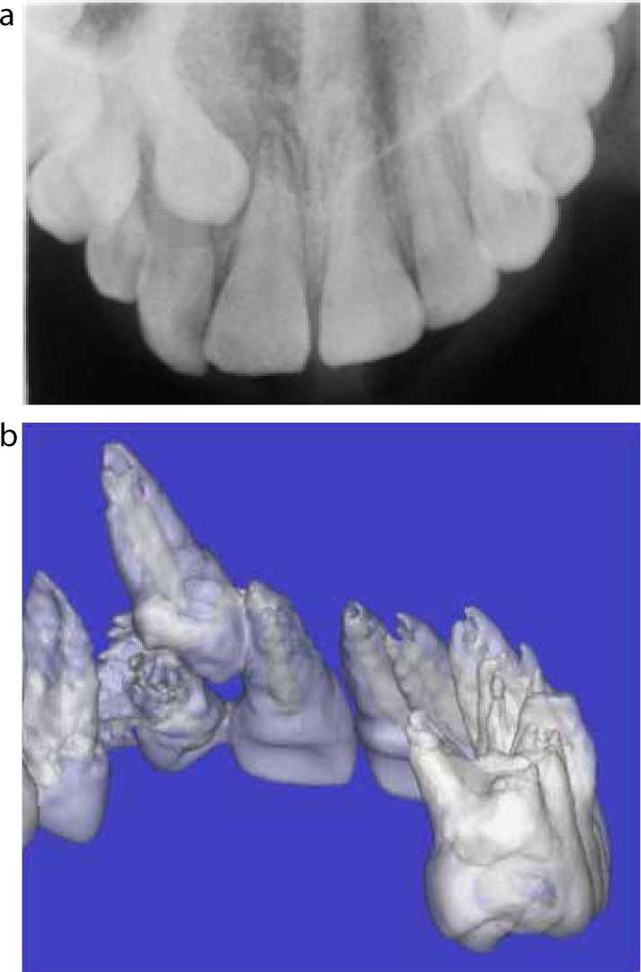
In addition to palpation, either buccally or palatally/lingually, there are other possible clinical indicators as to the likely position of the unerupted tooth. These include the position and mobility of adjacent teeth. Sometimes the inclination of the adjacent lateral incisor will indicate the position of the unerupted canine, particularly if the unerupted canine crown is pushing against the lateral incisor root (Figures 4 and 5). If the unerupted canine is not palpable, but the retained deciduous canine is mobile, the unerupted tooth is likely to be in a favourable position and probably close to eruption. Sometimes adjacent permanent teeth are mobile as a result of unwanted resorption caused by the ectopic canine.

Radiographic assessment of canine presence and position
The next step in identifying the position of the unerupted canine is a radiographic examination. The most common combinations of radiographs that can be used to locate an ectopically positioned maxillary canine are:
Of the two parallax techniques, horizontal parallax is reported to be more sensitive than vertical parallax.15
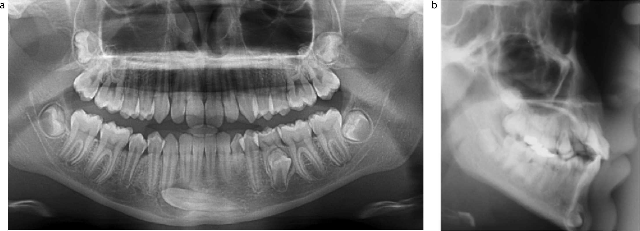
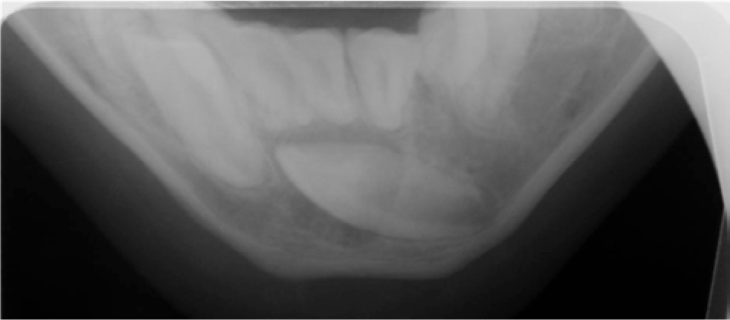
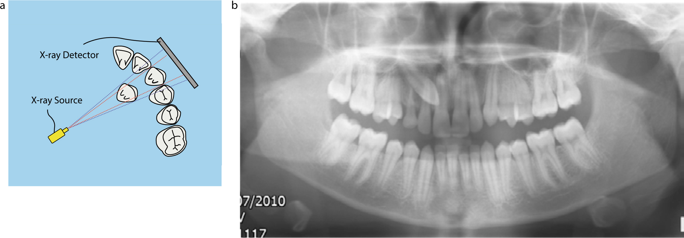
In addition to parallax, in the case of an ectopically positioned lower canine, a true 90° mandibular occlusal can sometimes be used on its own or two views at right angles. In orthodontics, a DPT and a lateral cephalometric view are commonly used for orthodontic diagnosis and, by chance, may also help localize an aberrant lower canine (Figure 6). Cone beam computed tomography (CBCT) can be used to locate ectopic canines accurately16 and identify possible resorption of the roots of the adjacent teeth3 (Figure 4), but this requires specialist equipment and will result in a higher radiation exposure to the patient. Standard lower occlusal radiographs (Figure 5) may not give sufficient information regarding the proximity of the canine crown to the lower incisor roots due to superimposition. Altering the X-ray angulation along the long axis of the lower incisors can give more diagnostic and accurate information (Figure 7). Another, less sensitive, method of assessing whether an ectopically positioned canine is palatally or buccally placed is based on its relative magnification on a single OPT radiograph.17 If the canine is palatally placed it will appear larger on the X-ray image, as it is closer to the X-ray source than the rest of the teeth in the arch (Figure 8).
Radiographic sector analysis of position and eruptive likelihood
When trying to predict the likelihood of success of interceptive treatments for the alignment of the ectopically positioned upper canine, a radiographic sector analysis can be used. Here the radiographic position of the canine crown is described as being in one of five sectors (Figure 9). The analysis was described by Ericson and Kurol18 and the five canine positions are:
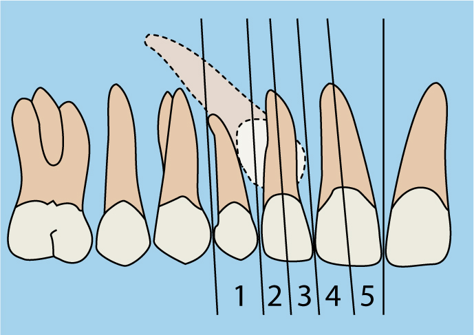
Both Ericson and Kurol18 and Power and Short19 reported that the more mesial the canine crown, the less the likelihood of successful interceptive treatment. Ericson and Kurol18 reported that, when the unerupted canine was in sectors 1 or 2, there was a 90% chance of correction following interceptive extraction, compared to a 64% chance if the unerupted canine was in sectors 3, 4 or 5, indicating the relationship between cusp tip and midpoint of the lateral incisor root to be a diagnostic predictor. This is a finding echoed by other researchers, who have suggested that the mesial position of the canine cusp tip is the most important factor in predicting the success of prospective interceptive treatment.20 However, it is not just the proximity of the unerupted upper canine to the midline that is important. The more parallel it is to, and the greater the distance it is from, the occlusal plane, the poorer the outcome of any interceptive extraction of the deciduous canine. Interceptive extractions will be discussed in part 2.
Conclusion
The eruption of the canine tooth is a significant dental milestone. Often this occurs without incidence, but the aberrant canine can be one of the most challenging treatment scenarios commonly faced by the orthodontist. Early detection can allow interceptive treatment to be performed and can significantly reduce the risk of adverse events, such as root resorption, and the complexity of definitive treatment. The clinician should be aware of the possible sequelae of unerupted canines. This paper has outlined the aetiology and diagnosis of the aberrant canine; part 2 will discuss the various management strategies.
