Article
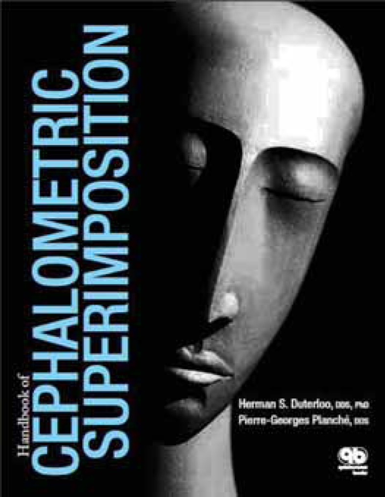
Handbook of Cephalometric Superimposition
This book is a study of the superimposition methods used in cephalometric radiography to quantify growth and the results of treatment. It takes the reader through the history of pre-radiographic methods of evaluating skull form and shape from the 18th Century, with an account of the work of John Hunter (1728–1793) in using madder dye to assess mandibular growth in pigs. Further developments are described, leading up to the work of the paleoanthropologist Sir Arthur Keith and the dentist George C Campion in 1922 in which they presciently superimposed their skull sections on the pituitary fossa and the cribiform plate – the two structures that were later found by Melsen to remain in a stable relationship to each other throughout growth.
The work of Arne Bjork is extensively referred to and the various methods of superimposition that he and his co-workers advocated are discussed in great detail. The advantages and disadvantages of each are also given, and the authors recommend the use of the ‘structural method’ of superimposition – on the basis that the traditional bone-based landmarks are unstable and change their relationship to one another with time and treatment (with the exception of adults over a short time period).
Other chapters are given to the problem of extremes of craniofacial growth, normal variations, and errors in superimposition, with the final chapter given over to a detailed account of how the structural method may be performed in both traditional handtracing and with the use of digital images on a desktop computer using Adobe Photoshop.
Such an approach may seem laudable, but the errors inherent in any cephalometric tracing are likely to make such an idealized approach impracticable. For example, the structural method relies on constructing an L-shaped template above the anterior cranial base and having the lower leg parallel to the Frankfort plane. The location of the Frankfort plane, in itself, is subject to errors of tracing and it is unlikely that most orthodontists will get it right every time.
The authors acknowledge that most current superimpositions can only give part of the picture of craniofacial growth, but for most orthodontic trainees, and for the majority of orthodontists, superimposition on the sella-nasion line, the maxillary plane, and the Bjork structures in the mandible are likely to continue to give the diagnostic information that is required.
As a text for those undergoing specialist training in orthodontics, this volume would be of somewhat limited use as a reference, in that most of what they need is covered elsewhere. As with all Quintessence books, the standard of presentation and illustration is high throughout, and anyone wishing to undertake research for a higher degree involving cephalometry will find it extremely useful.

