Isaacson KG, Thom AR, Horner K, Whaites E, 3rd edn. London: British Orthodontic Society; 2008
Whaites E, 4th edn. London: Churchill Livingstone Elsevier; 2007
London: HMSO; 2000
Manchester: European Commission; 2009
Recommendation of the International Commission on Radiological Protection. Ann ICRP. 2007; 37:1-332
Recommendation of the International Commission on Radiological Protection. Ann ICRP. 1991; 21:1-201
Danforth RA, Clark DE Effective dose from radiation absorbed during a panoramic examination with a new generation machine. Oral Surg Oral Med Oral Pathol Oral Radiol Endod. 2000; 89:236-243
Dula K, Mini R, van der Stelt PF, Buser D The radiographic assessment of implant patients: decision-making criteria. Int J Oral Maxillofac Implants. 2001; 16:80-89
Frederiksen NL, Benson BW, Sokolowski TW Effective dose and risk assessment from film tomography used for dental implant diagnostics. Dentomaxillofac Radiol. 1994; 23:123-127
Gori C, Rossi F, Stecco A, Villari N, Zatelli G Dose evaluation and quality criteria in dental radiology. Radiat Prot Dosimetry. 2000; 90:225-227
Lecomber AR, Downes SL, Mokhtari M, Faulkner K Optimisation of patient doses in programmable dental panoramic radiography. Dentomaxillofac Radiol. 2000; 29:107-112
Lecomber AR, Faulkner K Conference Proceeding: Dose and Risk in Dental Radiography, Luxembourg 1997. Reference doses and quality in medical imaging: what the referring practitioner and directing medical staff should know. Radiat Prot Dosimetry. 1998; 80:23-25
Pasler FA, Visser HAuflage: Georg Thieme; 1999
White SC Assessment of radiation risk from dental radiography. Dentomaxillofac Radiol. 1992; 21:118-126
Okano T, Harata Y, Sugihara Y, Sakaino R, Tsuchida R, Iwai K, Seki K, Araki K Absorbed and effective doses from cone beam volumetric imaging for implant planning. Dentomaxillofac Radiol. 2009; 38:79-85
Silva MA, Wolf U, Heinicke F, Bumann A, Visser H, Hirsch E Cone-beam computed tomography for routine orthodontic treatment planning: a radiation dose evaluation. Am J Orthod Dentofacial Orthop. 2008; 133:640.e1-e5
Palomo JM, Rao PS, Hans MG Influence of CBCT exposure conditions on radiation dose. Oral Surg Oral Med Oral Pathol Oral Radiol Endod. 2008; 105:773-782
Garcia Silva MA, Wolf U, Heinicke F, Gründler K, Visser H, Hirsch E Effective dosages for recording Veraviewepocs dental panoramic images: analog film, digital, and panoramic scout for CBCT. Oral Surg Oral Med Oral Pathol Oral Radiol Endod. 2008; 106:571-577
Gijbels F, Bou Serhal C, Willems G, Bosmans H, Sanderink G, Persoons M, Jacobs R Diagnostic yield of conventional and digital cephalometric images: a human cadaver study. Dentomaxillofac Radiol. 2001; 30:101-105
Maillie HD, Gilda JE Radiation-induced cancer risk in radiographic cephalometry. Oral Surg Oral Med Oral Pathol. 1993; 75:631-637
Visser H, Rodig T, Hermann KP Dose reduction by direct-digital cephalometric radiography. Angle Orthodontist. 2001; 71:159-163
Ludlow JB, Davies J, Ludlow LE, White SC Patient risk related to common dental radiographic examinations: the impact of 2007 International Commission on Radiological Protection Recommendations regarding dose calculation. J Am Dent Assoc. 2008; 139:(9)1237-1243
Watson SJ, Jones AL, Oatway WB, Hughes JJ Ionising Radiation Exposure of the UK Population Review 2005.
Joint Working Party of the British Society for the Study of Orthodontics and the British Society of Dental and Maxillofacial Radiology. The reduction of the dose to patients during lateral cephalometric radiography. Br J Orthod. 1985; 12:176-178
Gijbels F, Sanderink G, Wyatt J, Van Dam J, Nowal B, Jacobs R Radiation doses of collimated vs non collimated cephalometric exposures. Dentomaxillofac Radiol. 2003; 32:128-133
Svenson B, Sjoholm B, Jonsson B Radiation of absorbed doses to the thyroid gland in orthodontic treatment planning by reducing the area of irradiation. Swed Dent J. 2004; 28:137-147
Tsiklakis K, Donata C, Gavala S, Karayianna K, Kamenopoulou V, Hourdakis CJ Dose reduction in maxillofacial imaging using low dose Cone Beam CT. Eur J Radiol. 2005; 56:413-417
Senior Lecturer/Honorary Consultant in Dental and Maxillofacial Radiology, King's College London Dental Institute, Department of Dental and Maxillofacial Radiological Imaging, Floor 23, Tower Wing, Guy's Hospital, London, UK
This article provides an update on two-dimensional (2D) and three-dimensional (3D) imaging in contemporary orthodontics. The use of computed radiography (CR) and direct digital radiography (DR) for two-dimensional intra- and extra-oral imaging are outlined and compared. The advantages and disadvantages of each approach are discussed and various common imaging faults that may be produced are illustrated. 3D Cone-beam computed tomography (CBCT) image production is presented and discussed with consideration to patient dose and its applications in orthodontics. The current SEDENTEX CT guidelines for the appropriate use of this imaging modality are also outlined.
Clinical Relevance: As radiographic imaging is an integral aspect of clinical orthodontics understanding modern 2D and 3D imaging techniques is essential to maximize the benefits and minimize the risks to patients.
Article
Orthodontic treatment planning is primarily based on a thorough clinical examination, with radiographs being regarded as the orthodontist's main diagnostic aid. However, the range of radiographic imaging available, and how images are obtained, has changed considerably in recent years with the development of two-dimensional digital imaging and, more recently, with three-dimensional digital imaging. This article provides an overview of these digital imaging developments and their advantages and disadvantages as diagnostic aids in clinical orthodontics. Selection criteria for conventional radiographs in orthodontics will not be covered as these are available in the British Orthodontic Society's Guidelines for the Use of Radiographs in Clinical Orthodontics 3rd edition.1 However, brief reference is made to current guidance on the clinical use of 3D imaging.
2D digital imaging
Commonly used 2D orthodontic radiographs include:
Bitewings;
Periapicals;
Occlusals;
Panoramic radiographs;
Cephalometric lateral skulls.
Image acquisition for all of these views requires X-ray generating equipment (dental or panoramic) and some form of digital image receptor. The X-ray generating machines used in digital imaging are essentially identical to those that have been used traditionally in dentistry to create film-based images. The major development in imaging has resulted from changing the image receptor. Digital imaging receptors may be one of two types:
Photostimulable phosphor plates – used in what is commonly referred to as computed radiography (CR)
Solid state sensors – used in what is commonly referred to as direct digital radiography (DR).
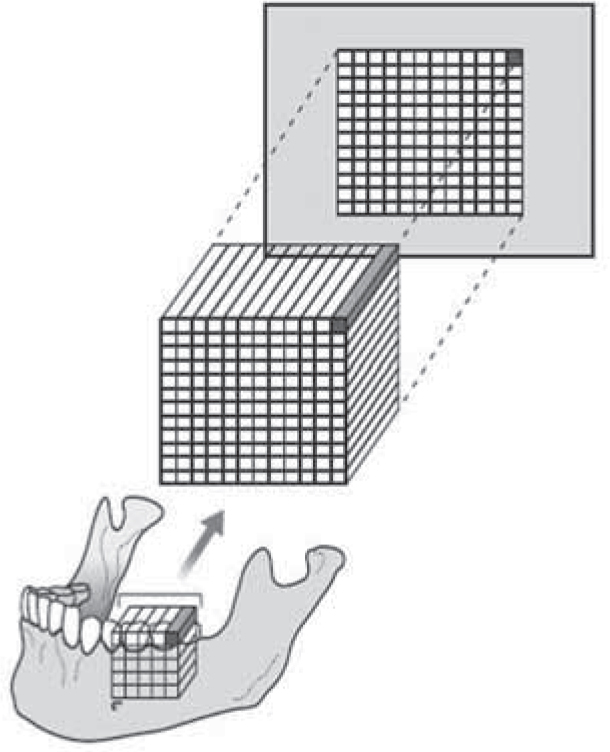
With both systems, the captured digital radiographic image is a two-dimensional representation of the three-dimensional patient and is made up of tiny black, white and grey square picture elements or pixels as shown in Figure 1. A typical image utilizes 256 shades of grey.
Figure 1. Diagram illustrating how the 3-dimensional jaw is represented as a digital image made up of a grid or a matrix of 2-dimensional pixels. (Reproduced from Whaites' Essentials of Dental Radiography and Radiology 4th edn with kind permission of Elsevier).2
Computed radiography using photostimulable phosphor plates
Computed radiography was developed by the Fuji imaging company, with the first medical Fuji Computed Radiography (FCR) system being released in 1982. The technology involved the use of photostimulable phosphor imaging plates which were constructed of europium barium fluorohalide. These original imaging plates were designed to replace the film and intensifying screens conventionally found in medical-sized cassettes. By the 1990s, dental-sized CR phosphor plates were available – 15 × 30 cm for panoramic radiography and 18 × 24 cm for lateral cephalometric radiography (Figure 2).
Figure 2.
(a) Original film-based lateral skull cassette containing light-sensitive film and two rare-earth light emitting screens and (b) modern CR lateral skull cassette with photostimulable phosphor plate.
Dedicated dental intra-oral phosphor imaging plates for bitewings and periapicals were first produced by the Soredex imaging company in 1994 for their Digora FMX system. However, the first dental computed radiography system to undertake occlusal imaging was the Gendex Denoptix system, which did not arrive on the market until 1999. Intra-oral phosphor plates are not used in cassettes but are individually wrapped in black paper and sealed within a plastic barrier envelope (Figure 3).
Figure 3. Original intra-oral film packet contents (top) and typical phosphor plate packaging (bottom).
There are currently four computed radiography systems dedicated to dental intra-oral and extra-oral imaging (Gendex, Soredex, Duerr, Kodak), while four medical imaging companies (Agfa, Fuji, Konica, Carestream) support dental extra-oral panoramic and lateral cephalometric-sized imaging plates.
In addition to photostimulable phosphor plates, CR systems also require a reader and a computer with (ideally) a medical grade image quality monitor. Following exposure of the plate to X-rays, the latent image is stored within the phosphor layer of the imaging plate, as a result of displacement of an electron within the atomic structure of the europium barium fluorohalide. The imaging plate is then placed within a reader where a laser beam is aimed at the plate. The laser causes re-alignment of the electrons and emission of energy in the form of light so that the imaging plate fluoresces. The emitted light is picked up by a photomultiplier tube within the reader and converted into an analogue voltage signal. This analogue signal is converted to a digital signal in the computer where the image is divided up into tiny square picture elements or pixels and each pixel is allocated a number – typically from 0 to 255, depending on the amount of X-ray radiation that reached each part of the plate. Finally, each pixel is allocated a shade of grey from black through to white and the radiographic image is displayed on the computer monitor. The phosphor plate is finally flooded by ambient light to erase the image, allowing the plate to be re-used for further imaging.
Advantages of computed radiography (CR)
Conventional X-ray generating equipment does not need to be replaced, however, liaison with a medical physicist may be appropriate to adjust the exposure for optimization of the image.
Extra-oral cassettes containing film and intensifying screens are simply replaced by cassettes, only containing phosphor plates.
Intra-oral bitewing and periapical techniques are identical to conventional techniques using film packets.
Conventional intra-oral film packet holders with beam-aiming devices can still be used, making clinical usage straightforward.
Phosphor plates have a wide latitude, ie can tolerate small (under-exposed) or large (over-exposed) amounts of radiation, but still enable a diagnostically useful image to be displayed following image manipulation.
Images can be viewed on computer monitors, printed on to paper or burnt to digital film.
Disadvantages of computed radiography (CR)
Images are not displayed instantaneously – phosphor plates have to be read, although scanning time is nowadays typically less than 10 seconds for intra-oral views, and 60 seconds for extra-oral images.
Wide latitude means less attention can inadvertently be paid to ‘exposure factors’, but patients may be unnecessarily over-exposed. All exposures should still be kept as low as reasonably practicable.
Phosphor crystals are receptive on both sides so that plates can be exposed back to front and still produce an acceptable image (Figure 4a).
Some of the dedicated dental imaging systems do not automatically erase the imaging plate post-examination and require the operator to flood the plate with ambient light to ensure it has been erased.
As ambient light causes erasure of the image, readers should be positioned in rooms with reduced or dimmed lighting (Figure 4b).
A number of the dental imaging systems require considerable handling of the phosphor plates (wrapping and unwrapping) thereby putting the plates at risk of damage from the operator, eg scratching (Figure 4c).
Only the dental manufacturers provide dental intra-oral imaging plates that can only be read within dedicated readers.
Many manufacturers only provide Size 2 and Size 4 imaging plates that are exactly the same size as dental film. The Size 0 imaging plates are shorter and the Size 1 plates longer than the equivalent matched-sized film!
As occlusal radiography requires the patient to bite gently on to the image receptor, occlusal phosphor plates must be protected from mechanical damage during clinical use (eg with perspex sheets as shown in Figure 5).
Figure 4. Imaging plate faults: (a) plate exposed back-to-front; (b) plate exposed to bright ambient light causing partial erasure; and (c) scratched plate.Figure 5. Occlusal image plate protection from mechanical damage using paper covers and perspex ‘bite’ sheets.
Direct digital radiography (DR) using solid state sensors
Direct digital radiography was first introduced to the medical imaging market via dentistry when the first direct digital imaging sensors were released by Trophy imaging in 1987 as the RadioVisioGraphy (RVG) imaging system. Since then, many further direct imaging sensors have been released for both dental (intra-oral and extra-oral) and medical imaging using two main types of solid state detectors:
Charged couple devices (CCD); and
Complementary Metal Oxide Semiconductors (CMOS).
Intra-oral sensors
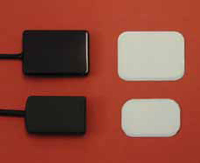
Currently, these imaging receptors are available in two sizes for intra-oral bitewing and periapical radiography (usually adult and child size), however, as shown in Figure 6, these are slightly smaller than equivalent dental film packets and the plastic sensor casing is commonly larger than the receptive area for imaging. In most systems, the sensors are directly connected to the computer by a cable or wire, although currently a number of manufacturers also offer a wireless imaging option. Unfortunately for orthodontists, occlusal-sized sensors are not available.
Figure 6. Intra-oral child- and adult-sized solid-state sensors and equivalent-sized film packets.
As shown in Figure 7, the CCD sensors are made up of individual pixels, consisting of a sandwich of P- and N-type silicon, arranged in rows and columns called an array or matrix, above which is a scintillation layer made of similar materials to rare-earth intensifying screens.
Figure 7. Diagrams illustrating the basic construction of a CCD sensor. (A) The imaging surface showing the pixel array; (B) sensor from the side showing the scintillation layer; and (C) an individual pixel consisting of a sandwich of P- and N-type silicon. (Reproduced from Whaites' Essential of Dental Radiography and Radiology 4th edn with kind permission of Elsevier).2
The clinical image is created by the X-ray photons, that have passed through the patient, striking the scintillation layer which absorbs and converts them into visible light. The light interacts with the silicon to create a charge packet for each individual pixel, which is concentrated by the electrodes. The charge pattern formed from the individual pixels in the matrix represents the latent image. The image is read by transferring each row of pixel charges from one row to the next. At the end of each of its rows, each charge is transferred down the cable as an analogue voltage signal to the computer's analogue to digital converter, often located in a docking station. Each sensor consists of 1.5–2.5 million pixels. As with CR systems, each pixel is allocated a number – typically 0–255, dependent on the amount of radiation received, then allocated a grey-scale colour to produce the black, white and grey radiographic digital image on the monitor. The images are produced instantaneously and, unlike computed radiography, no reader or erasure of the old image is required, so the sensor is immediately ready to be used again clinically to produce another image.
Extra-oral sensors
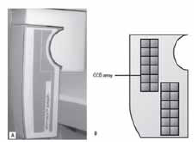
This technology has been adapted for use in larger field medical and extra-oral dental imaging by utilizing a thin, narrow X-ray beam aligned to a thin, narrow linear CCD array (or arrays) which together scan across the region of interest to capture the imaging data. As shown in Figure 8, if more than one CCD array is used, a larger imaging sensor can be created. The captured data is then stitched together to produce one large image; or is, alternatively, achieved by a method called optical coupling.
Figure 8.
(A) specifically designed Planmeca dimax solid state sensor for panoramic and cephalometric radiography. (B) Diagram showing the two thin internal CCD arrays. The lower array of CCDs are used to construct the panoramic image. The upper and lower arrays are both used in combination to construct a cephalometric image by stitching the two separate datasets together. (Reproduced from Whaites' Essentials of Dental Radiography and Radiology 4th edn with kind permission of Elsevier.)2
As conventional panoramic radiography already uses a thin, narrow X-ray beam that scans/rotates around a patient's head, it lends itself to CCD technology and the use of a thin, narrow CCD array as the image receptor. The first commercial CCD-based direct digital dental panoramic machine was developed by Siemens in 1997. As the panoramic imaging is built up incrementally, the direct digital image is displayed instantly, but incrementally, on the computer screen.
CCD-based scanning technology was also developed for lateral skull radiography. Again Siemens introduced the first system which relied on the sensor and the thin, narrow X-ray beam scanning in a vertical direction, moving from superior to inferior and capturing the full image of the facial skeleton incrementally. The same sensor could be used to capture the panoramic image and cephalometric image, but needed to be transferred into a different sensor holder between techniques. This concept has been superseded by most current systems employing the sensor and the thin, narrow X-ray beam scanning in a horizontal direction, moving from anterior to posterior as shown in Figure 9. To ensure exact alignment of the X-ray beam with the sensor, most equipment includes a secondary collimator which also moves during the exposure. Horizontal scanning has the advantage of enabling larger areas than the typical 18 × 24 cm to be imaged (potentially up to 30 × 24 cm) and there is less magnification.
Figure 9. Close-up of the Planmeca Cephalostat: (a) at the start of the exposure; and (b) at the end. This equipment is designed to scan the patient horizontally with the X-ray beam, secondary collimator and sensor moving horizontally throughout the exposure (arrowed). (Reproduced from Whaites' Essentials of Dental Radiography and Radiology 4th edn with kind permission of Elsevier.)2
Currently, two manufacturers have developed direct digital imaging receptors for lateral skull radiography that allow image capture in a so-called one shot technique, as in conventional film-based imaging, without scanning:
Kodak Dental Imaging uses a CCD image receptor and a method of optical coupling; and
Vatech Ewoo imaging utilizes a single thin flat panel imaging detector.
Advantages of direct digital radiography (DR)
Image capture is instantaneous.
Operator handling of intra-oral sensors is minimal and almost non-existent for extra-oral sensors.
Radiation dose is potentially less than film-based imaging.
Images can be viewed on computer monitors, printed on to paper or burnt to digital film.
Disadvantages of direct digital radiography (DR)
Intra-oral dental sensors are completely rigid and relatively bulky which can cause clinical difficulties in accurate sensor placement.
Imaging area of intra-oral dental sensors is smaller than conventional films so the area of interest may be missed.
The connecting cable may cause additional clinical problems in accurate sensor placement.
Most intra-oral sensors and cables are not autoclavable/sterilizable so need to be protected from salivary contamination with plastic barrier coverings.
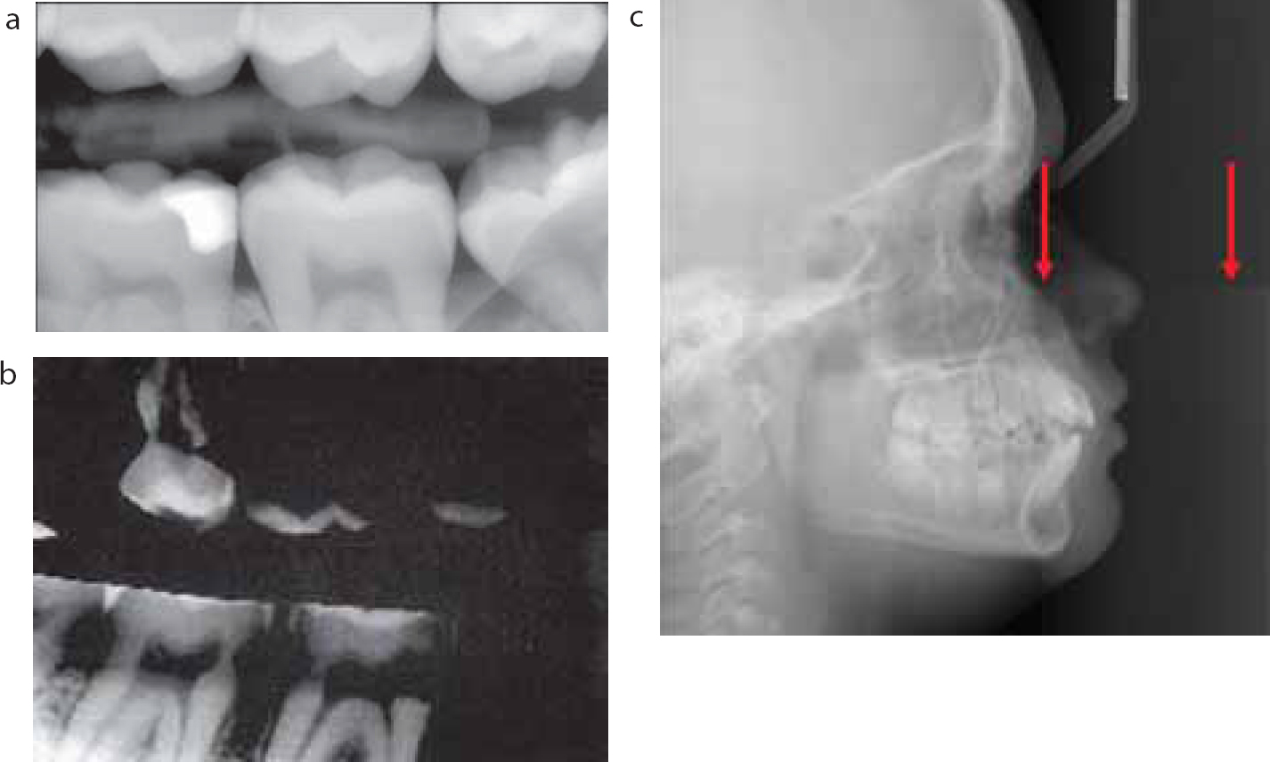
If dropped, the sensor can crack, producing a permanent image artefact (Figure 10a).
Occlusal-sized, solid-state sensors are not manufactured.
Patients have to remain still for several seconds during an exposure when using extra-oral equipment employing a scanning motion similar to plain film imaging.
Sensor latitude is not as great as for phosphor plates and overexposure can lead to CCD overloading or ‘blooming’ (Figure 10b).
Stitching of different scans together can produce artefacts on the final large composite image (Figure 10c).
Previously recommended triangular collimation of the X-ray beam during cephalometric radiography, which avoided unnecessary irradiation of the calvarium and cervical spine, has been lost.
Figure 10. Solid state sensor artefacts: (a) cracked scintillator screen; (b) blooming; and (c) stitching artefact.
Three dimensional imaging
Conventional, film-based imaging and the digital imaging described above share one fundamental limitation – both result in 2-dimensional images of the 3-dimensional patient, as illustrated in Figure 1. In recent years, there has been an ever growing demand in all branches of medicine to be able to see the missing third dimension. In dentistry, the main driver has been implantology. Historically, this third dimension has been imaged using medical computed tomography (CT) but, because of the high radiation dose involved, its use in orthodontics has been mainly limited to complex craniofacial problems. During the last 10 years, cone-beam computed tomography (CBCT) – three-dimensional digital imaging – for dental use has been developed and refined with potential applications in all branches of dentistry.
Cone-beam CT
Cone-beam computed tomography sometimes, and perhaps more accurately, referred to as cone-beam volumetric imaging, involves a conventional cone-shaped X-ray beam and an imaging detector. The object to be scanned is imaged as the radiation source rotates around the stationary object and irradiates the two-dimensional image detector, thereby capturing essentially a number of 2D views of the object from 360 different positions. This allows a single rotation of the radiation source to capture the volume of interest. These multiple 2D views are then amalgamated into a single cylindrical or spherical dataset, made up of tiny cubes or voxels of data. As with 2-dimensional pixels, each voxel is allocated a number and then allocated a shade of grey from black through to white. The computer then allows the dataset (the voxels) to be re-arranged and viewed as conventional axial, sagittal and coronal images as shown in Figure 11.
Figure 11. Diagram showing the basic concept of cone-beam CT. A cone-shaped X-ray beam is used which rotates around the patient obtaining information in a volume. The patients maxillofacial skeleton is positioned within the region of interest and is divided into tiny cubes or voxels. Computer manipulation (multiplanar reconstruction) of the data obtained allows separate images in the sagittal, coronal and axial planes to be created. (Reproduced from Whaites' Essentials of Dental Radiography and Radiology 4th edn with kind permission of Elsevier.)2
In addition, using the imaging dataset, the voxels may be reconstructed into panoramic and lateral cephalometric images, as well as axial images similar to occlusal views. Other images, such as cross-arch, airway views and three-dimensional models, may also be obtained from the dataset.
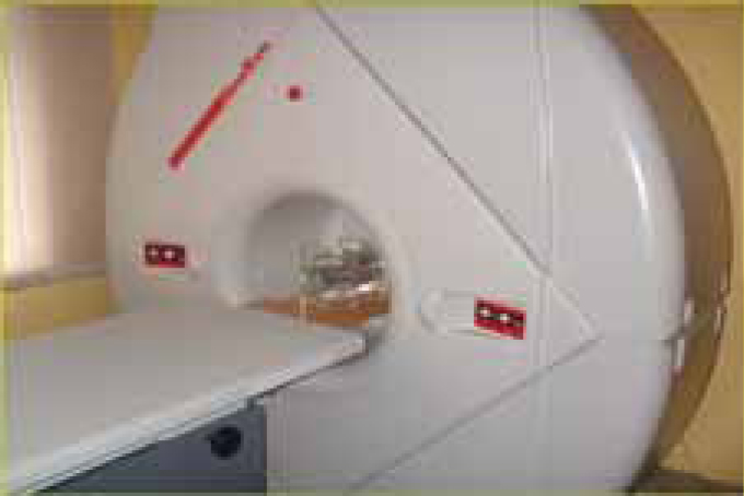
The first CBCT system released on the dental market was the Newtom 9000 shown in Figure 12. This unit resembled a medical CT scanner. The patient lay supine on an imaging table, and his/her head was placed within a scanning unit. The X-ray source and imaging detector made a 360 degree revolution around the head and captured individual images at each degree of rotation. The original image detectors were image intensifiers, which have been available since the 1970s and used extensively in medical fluoroscopy units. Image intensifiers convert X-rays into a light signal which is then read by a CCD detector. The signal is then sent to a computer and displayed as a digital image as described earlier. Owing to the round shape of an image intensifier, the imaging volume collected is normally spherical.
Figure 12. The original Newtom CBCT units which utilized an image intensifier as image detector. (Kindly provided by Mr C Cook).
Almost all the second generation of CBCT units utilize rectangular flat panel detectors as the image receptor. These flat panels enable a cylinder of 3D imaging data to be captured. They can also be used for conventional 2D digital imaging. Hence some modern CBCT units can take both conventional panoramic and CBCT images. Flat panel detectors are, however, very expensive to replace and they require a higher radiation dose. However, the flat panel detector systems are typically smaller than an image intensifier-based imaging system and therefore require less physical space. Some CBCT units currently undertake only a 180 or 270 degree rotation, which results in a half to two thirds of the number of 2D frames captured compared to a unit making a 360 degree rotation. This has the potential to decrease the patient dose and still produce three-dimensional images but, as less raw data is being initially collected in these scans, the final dataset may be more susceptible to artefacts than those undertaking a full rotation.
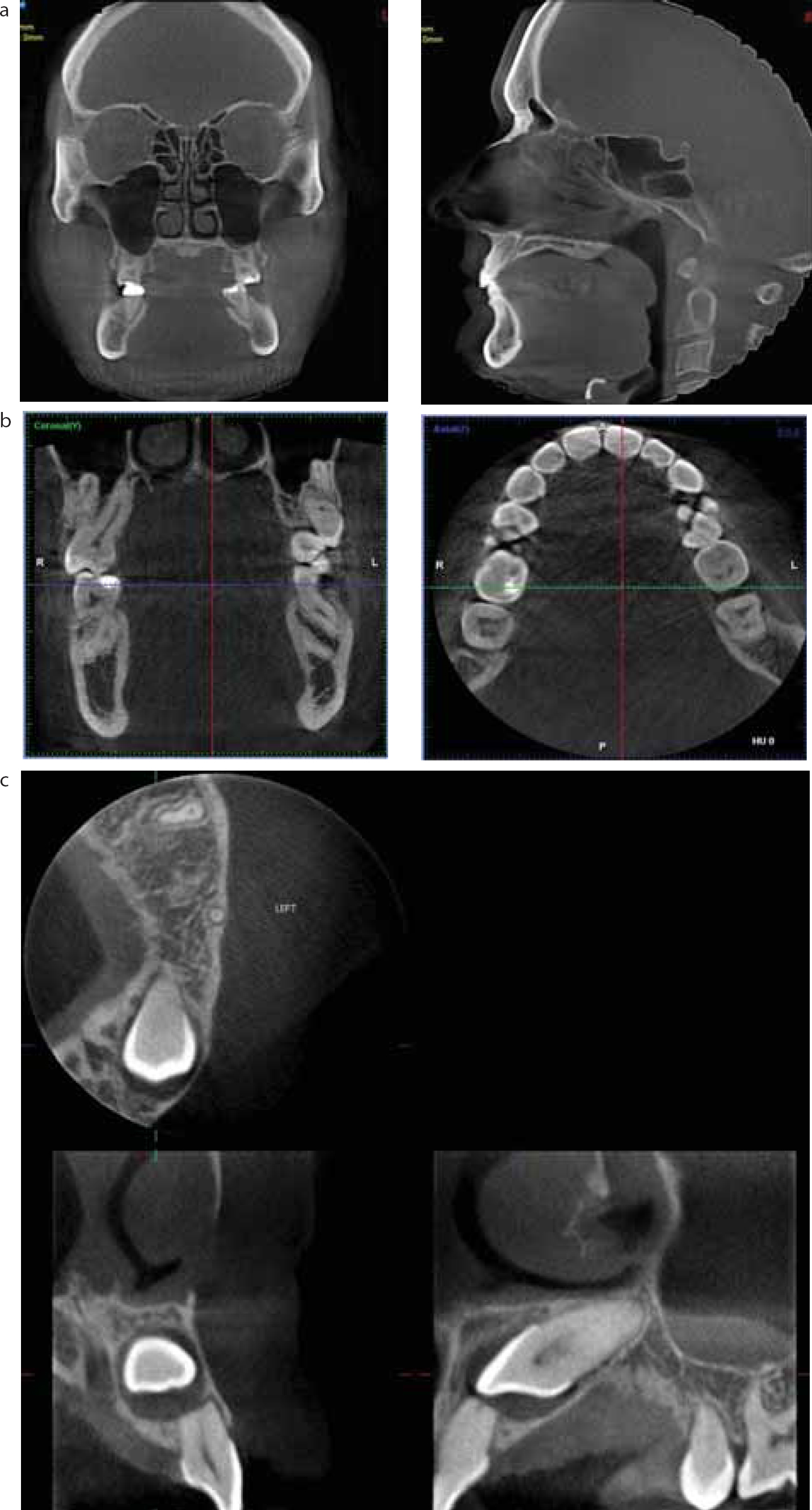
The major difference in the approximately 15–20 CBCT units currently available on the market is in the size of their ‘field of view’ (FOV), as shown in Figure 13. Essentially these divide into:
Large FOV – eg 23 cm × 26 cm capable of imaging most, if not all, of the head;
Medium FOV – eg 8 cm × 8 cm capable of imaging the mandible and maxilla (the jaws);
Small FOV – eg 4 cm × 4 cm capable of imaging just 2–3 individual teeth.
Figure 13. Examples of CBCT images with different-sized fields of view captured by different CBCT units: (a) large field of view approximately 30 cm high showing the full maxillofacial skeleton and cranium as coronal and sagittal images; (b) medium field of view limited to the mandible and maxilla as coronal and axial images; (c) small field of view showing just the left anterior teeth as axial, coronal and sagittal images.
Most units are now capable of offering practitioners a series of different-sized fields of view – an important consideration in orthodontics.
Clinical applications of CBCT in orthodontics
There is no doubt that these CBCT units result in exquisite images, enabling the missing third dimension to be visualized, but the radiation dose involved can be considerably higher than from conventional film-based or 2D digital imaging. Therefore, the key question to be asked is ‘Is 3D radiographic information going to affect patient management?’. In compliance with The Ionising Radiation (Medical Exposure) Regulations 2000,3all medical exposures, including CBCT, have to be justified. As stated initially, orthodontic treatment planning is primarily based on a thorough clinical examination of a 3-dimensional patient. If any additional information is required, can the answer be provided by low dose 2D conventional imaging? If not, then 3D CBCT may be required.
At the present time, no research evidence base exists from which to develop evidence-based guidelines on the use of CBCT in orthodontics. The Provisional SEDENTEX CT European Guidance (May 2009)4 states:
The applications of CBCT in assessment of the developing dentition for orthodontics will be considered under two broad headings: localised application to answer a specific question and generalised application for the examination of the entire dento-facial region.
Applications in orthodontics
Conventional radiography has served orthodontists well over many years and, at the present time, no research evidence base exists from which to develop evidence-based guidelines on the use of CBCT in orthodontics. The current European-wide SEDENTEX CT project's Guideline Development Panel concluded in their Provisional Guidance, published in May 2009, that there is a need for research demonstrating change (and improved) management of patients before routine use of CBCT for orthodontics could be considered.4
The SEDENTEX project considered CBCT applications in the assessment of the developing dentition for orthodontics under two headings: localized (small volume/FOV) application to answer a specific question and generalized (large volume/FOV) for the examination of the entire dento-facial region and proposed that the following clinical applications may be appropriate for CBCT usage:
For the localized assessment of an impacted tooth (including consideration of resorption of an adjacent tooth), where the current imaging method is conventional medical CT, CBCT may be preferred because of reduced radiation dose.
For the localized assessment of an impacted tooth (including consideration of resorption of an adjacent tooth) where the current imaging method of choice is conventional dental radiography, CBCT may be used when the information cannot be obtained adequately by lower dose conventional radiography.
Where the current imaging method of choice for the assessment of cleft palate is conventional medical CT. CBCT may be preferred where the radiation dose is lower. The smallest volume size compatible with the situation should be selected because of reduced radiation dose.
Large volume CBCT should not be used routinely for orthodontic diagnosis.
For complex cases of skeletal abnormality, particularly those requiring combined orthodontic/surgical management, large volume CBCT may be justified in planning the definitive procedure, particularly where conventional medical CT is the current imaging method of choice.
Research is needed to define robust guidance on clinical selection for large volume CBCT in orthodontics, based upon quantification of benefit to patient outcome.
Disadvantages
Dose and risk considerations
The most useful radiation dose measurement that allows comparisons to be made between different radiographic investigations is the effective dose. Effective dose takes into account the following:
The amount of radiation energy absorbed;
The damaging effect of the type of radiation being used; and
The sensitivity of the tissues being irradiated.
The International Commission on Radiological Protection (ICRP) periodically publishes the individual tissue weighting factors and, in 20075, published the current weighting factors which replaced their 19906 figures. These new tissue weighting factors include a specific weighting for the salivary glands and overall increase the effective doses from dental radiography.
Individual effective doses in 2D digital dental radiography (intra-oral, panoramic and cephalometric) are low, being equivalent to those associated with a few days of background radiation. In general, there is little difference in the dose for digital panoramic and cephalometric images compared with their film-based counterparts. Individual doses from more complex imaging, such as 3D CBCT scans can, however, be substantially higher and hence carry a greater risk.
Published effective doses for 2D digital radiographs vary considerably, depending on the type of equipment being used.
Panoramic
Using the ICRP 1990 weighting factors – 3.85–30 μSv;7,8,9,10,11,12,13,14
Using the ICRP 2007 weighting factors – 2.7–23 μSv.15,16,17,18
Cephalometric lateral skull
Using the 1990 weighting factors – 2–3 μSv;19,20,21
Using the ICRP 2007 weighting factor – 2.2–5.6 μSv.22
Therefore, using the latest ICRP weighting factors, a typical panoramic and cephalometric orthodontic examination will expose the patient to an effective dose in the range of 4.9 to 28.6 μSv. This compares with an average annual natural (not including medical imaging or consumer products) background radiation dose in the United Kingdom of 2.7 μSv.23 Although the radiographic examination dose is only a small fraction of natural background radiation, it is of course in addition to natural background.
These doses can be considerably reduced by employing:
Field limitation techniques for panoramic radiography, particularly by using the so-called ‘dentition only’ programme as shown in Figure 14. Reducing the field size by 40%, to limit the radiation field to the dentition only, achieves a 50% reduction in effective dose to the patient.11 The resultant smaller image often answers the clinical question required from the radiographic examination, particularly in orthodontics.
Triangular collimation for cephalometric radiography which reduces the irradiated field size by 55%24 by eliminating most of the cranial vault and cervical vertebrae. Unfortunately, as already mentioned, this technique has not yet been adapted for use with modern dental imaging equipment, although experimentally it has been shown to give an approximate 47% reduction in effective dose.25 However, this is based on the 1990 ICRP tissue weighting factors.
A thyroid shield/collar for occlusal radiography which protects the thyroid gland from the downwardly angulated X-ray beam.26
Figure 14. A full dental panoramic radiograph, showing the area depicted when using the ‘dentition only’ programme. The smaller image results in a 50% dose reduction.
There are a number of factors that affect the radiation dose delivered by 3D digital CBCT systems. These include:
Imaging parameters used (kVp, mAs);
Voxel size (resolution);
Pulsed or continuous radiation beam;
Amount, type and shape of beam filter;
Extent of rotation (full 360° rotation or less);
Size of the field of view.
Some of these factors, such as type of beam and filtration, are unique to individual machines, while other factors, such as field of view size, are under the control of the operator.
In general, the smaller the field of view, the lower the radiation dose. Since the effective dose is computed from a weighted summation of doses to various organs/tissues, removing some organs/tissues from the path of the X-ray beam will obviously reduce the effective dose to the patient. For example, since the radiation received by the thyroid gland contributes a large amount to the effective dose, limiting the beam and field of view to the maxilla and mandible and avoiding the neck results in a lower effective dose. As with occlusal radiography, the dose to the thyroid gland may also be reduced by tilting the head or using thyroid collars.27
Variable risk estimates of fatal cancer induction from dental radiography have been published over the years. An approximate summary of these are:
Dental panoramic radiograph – 1 in 1 000 000;
Lateral cephalometric radiograph – 1 in 5 000 000;
Cone Beam CT examination – 1 in 20 000 to 1 in 2 000 000 (wide variation given wide dose differences).
Large field of view CBCT investigations should therefore not be used as a direct replacement for the traditional orthodontic panoramic and cephalometric examinations. It is also not appropriate to take large field of view CBCT examinations solely to reconstruct panoramic or cephalometric radiographic views. The increased risk cannot be justified.
Conclusion
Radiographic investigations remain the orthodontist's main diagnostic aid. The relentless move from film-based imaging to 2D digital imaging is gathering momentum and offers clinicians many advantages, but disadvantages still exist. Caution needs to be exercised when considering 3D CBCT imaging in orthodontics. Although very helpful in solving specific clinical problems in orthodontics, there is no evidence base, as of yet, to justify its use routinely.