References

Morea C, Dominguez GC, Wuo ADV, Tortamano A Surgical guide for optimal positioning of mini implants. J Clin Orthod. 2005; 39:317-321
Cousley RRJ, Parberry DJ Surgical stents for accurate miniscrew insertion. J Clin Orthod. 2006; 40:412-417
Park HS, Jeong SH, Kwon OW Factors affecting the clinical success of screw implants used as orthodontic anchorage. Am J Orthod. 2006; 130:18-25
Estelita CBS, Janson G, Chiqueto K, de Freitas MR, Henriques JF, Pinzan AA Three-dimensional radiographic-surgical guide for mini-implant placement. J Clin Orthod. 2006; 40:548-554

Tricks of the trade: barium marker - a simplified guide for micro-implant placement

From Volume 8, Issue 3, July 2015 | Pages 108-109

Authors

Dharma RM

Professor, Department of Orthodontics and Dentofacial and Orthopaedics

Articles by Dharma RM

Dinesh MR

Professor and HOD, Department of Orthodontics and Dentofacial and Orthopaedics

Articles by Dinesh MR

Prashanth CS

Professor, Department of Orthodontics and Dentofacial and Orthopaedics

Articles by Prashanth CS

Amarnath BC

Professor, Department of Orthodontics and Dentofacial and Orthopaedics, DAPMRV Dental College, No CA 37, 24th Main, 1st Phase, JP Nagar, Bangalore, Karnataka, India-560 078

Articles by Amarnath BC

Article

Micro-implants are a contemporary source of anchorage in difficult situations and the accurate placement of these micro-implants is the cornerstone of a successful treatment plan.

Optimal positioning has always been critical to the effectiveness of dental implants. The choice of location depends on the initial diagnosis, the purpose of the implant therapy, the proximity of adjacent structures, such as the mandibular nerve and maxillary sinus, and aesthetic factors.1

Although orthodontic micro-implants require a less complex surgical procedure than normal implants, the quantity of interproximal bone and the inclination and proximity of the adjacent tooth roots need to be evaluated, otherwise there is a risk of root perforation.1,2,3

A careful clinical and radiographic assessment before implant placement is therefore a necessity.3 In this article, the authors suggest the use of barium sulphate for radiographic localization of micro-implant placement position.

Barium sulphate is frequently used clinically as a radiological contrast agent for X-ray imaging and other diagnostic procedures. It is most often used in imaging of the GI tract. This can be made use of in micro-implant placement together with crystal violet or gentian violet, which is a histological stain. Crystal violet has antibacterial, antifungal, and antihelminthic properties and was formerly important as a topical antiseptic. It can be used for marking the attached gingiva to facilitate micro-implant insertion.

The authors have developed a simple, reliable and accurate method for placing micro-implants as a single-step procedure.

Procedure

Before placement of the micro-implant, radiographic localization of the area is undertaken:
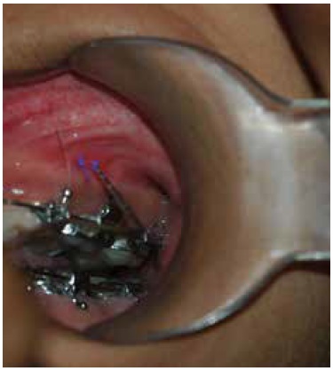
  • Thoroughly dry the area where the implant is intended to be placed (Figure 1).
  • Mark the interdental area with a periodontal probe along the long axis and also at the approximate area of placement (Figure 2).
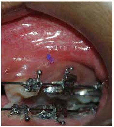
  • Mix barium sulphate with gentian violet into a thick ‘milk shake’ consistency and form a small ball which can be placed on the attached gingiva in the area located by the probe (Figure 3).
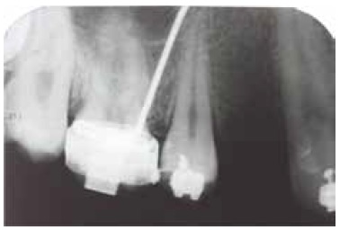
  • An intra-oral periapical radiograph (IOPA) of the area is taken at 0° angulation or bitewing angulation (Figure 4).
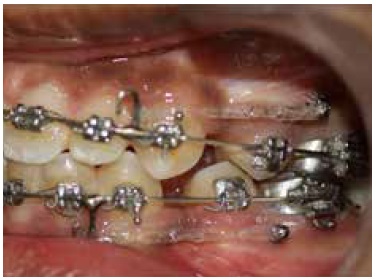
  • The IOPA is assessed to confirm the exact location for placement of the implant, which is shown by the radio-opacity of the barium marker. The implant is placed using the part of the attached gingiva stained by gentian violet as the clinical reference (Figure 5).
  • Figure 1. The area where the implant is intended to be placed.
    Figure 2. The approximate interdental area of implant placement where a mark is made using a periodontal probe along the long axis.
    Figure 3. A mark of barium sulphate with gentian violet in the approximate interdental area of implant placement.
    Figure 4. The IOPA with radio-opaque area.
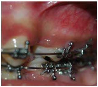
    Figure 5. A clinical view after successful implant placement.

    Discussion

    Various authors have proposed determining the implant position by utilizing customized surgical stents.4 Preparation of these stents can be time consuming and cumbersome. In contrast, the procedure described above has inherent advantages which include less chairside time and a simple, easy technique. The procedure can be repeated if the placement position has to be refined. The only potential disadvantage is that the angulation of placement of implant cannot be defined in the technique.