Sharifi A, Jones R, Ayoub AF, Moos KF, Walker FS, Khambay BS, McHugh S. How accurate is model planning for orthognathic surgery?. Int J Oral Maxillofac Surg. 2008; 37:1089-1093
Barbenel JC, Paul PE, Khambay BS, Walker FS, Moos KF, Ayoub AF. Errors in orthognathic surgery planning: the effect of inaccurate model orientation. Int J Oral Maxillofac Surg. 2010; 39:1103-1108
O'Malley AM, Milosevic A. Comparison of three facebow/semi-adjustable articulator systems for planning orthognathic surgery. Br J Oral Maxillofac Surg. 2000; 38:185-190
Tamburrino RK, Linton J. The axis-horizontal reference line for precision diagnosis: Part 1. Orthod Pract US. 2012; 3:25-28
Pitchford JH. A re-evaluation of the axis-orbital plane and the use of orbitale in a facebow transfer record. J Prosthet Dent. 1991; 66:349-355
Moorrees CFA, Kean R. Natural head position: a basic consideration in the interpretation of cephalometric radiographs. Am J Phys Anthrop. 1958; 16:213-234
Chan CA. A review of the clinical significance of the occlusal plane: its variation and effect on head posture. International College of Craniomandibular Orthopedics (ICCMO) Anthology. 2007; 8:1-63
Walker FS, Ayoub AF, Moos KF, Barbenel J. Face bow and articulator for planning orthognathic surgery: 1 face bow. Br J Oral Maxillofac Surg. 2008; 46:567-572
Walker FS, Ayoub AF, Moos KF, Barbenel J. Face bow and articulator for planning orthognathic surgery: 2 articulator. Br J Oral Maxillofac Surg. 2008; 46:573-578
Orthognathic surgery involving the maxilla generally requires the mounting of models on an articulator. Mounted casts facilitate detailed patient assessment and treatment planning, as well as allowing wafer construction to aid the three-dimensional surgical correction of dento-facial deformities and malocclusion. Systematic error in facebow transfer from the patient to the articulator is a contributing factor in the discrepancy between planned surgical movements and the final surgical result. This paper presents a simple technique that uses mini-spirit levels to allow commercially available semi-adjustable articulators to allow natural head position to be used as the horizontal reference plane rather than anatomical landmarks. We feel that this technique is another step towards error reduction in orthognathic surgery.
Clinical Relevance: Many orthognathic centres use commercially available semi-adjustable articulators that use anatomical landmarks to record the horizontal reference plane during facebow recording. These horizontal reference lines can differ significantly from the true horizontal line on which the patient is planned. The cross member of the articulator corresponds to the horizontal reference plane recorded and, if this is different from the true horizontal, then systematic error is introduced into orthognathic surgery.
Article
Orthognathic surgery involving the maxilla generally requires the mounting of models on an articulator. Mounted casts facilitate detailed assessment of the occlusion and allow model surgery to confirm the orthognathic plan prescribed by the clinical team. The mounted casts also facilitate construction of surgical wafers to aid the three-dimensional surgical movements to correct dento-facial deformities and malocclusion.1 The aim of this article is to inform the reader of one of the main potential sources of error in treating orthognathic patients, the transfer of maxillary position from patient to articulator. The authors also describe a simple method of facebow recording that reduces inaccuracies in the roll and pitch of the mounted maxillary cast.
A facebow is an instrument that uses three-point localization by two posterior references approximating to each of the temporomandibular joints, and an anterior reference point to relate the maxillary cast vertically to the selected horizontal reference plane.2 The purpose of this is two-fold:
To relate the maxillary arch to the condylar hinge axis of the mandible in three planes of space; and
To position the maxillary cast on the articulator as it was when the patient was planned for surgery.
The facebow recording and subsequent mounting of models is a potential source of error.1 If the pitch, roll or yaw of the models is inaccurate, this will mean that the model surgery movements will differ from the planned surgical movements, even if the laboratory prescription has been followed precisely.2 It has been proposed that altering the pitch of the mounted models also changes the estimation of autorotation during model surgery.3 This latter source of error could lead to the under- or over-estimation of mandibular movement required in a bimaxillary osteotomy or the amount of maxillary advancement required in a maxillary only osteotomy.
Various antero-posterior reference planes can be used to position the patient during assessment and align the outer bow of the facebow when recording maxillary position.4 These reference lines include:
The Frankfort horizontal plane;
The axis-orbitale plane;
Campers plane; and
A plane connecting the external auditory canal with an arbritary point relative to the tip of the maxillary lateral incisor.
Using orbitale as the anterior reference point has been shown to produce mounting errors of maxillary casts.5
The clinician can choose to position the patient in natural head position when assessing and planning for treatment. Natural head position was defined by Broca (1862) as ‘when a man is standing and when his visual axis is horizontal, he is in natural position’. One most widely used method for adopting a reproducible natural head position was described by Moorrees and Kean,6 where subjects were instructed to sit upright on a stool and asked to look into the image of their eye in a mirror located at the same level as the pupils of their eyes. The use of natural head position in conjunction with the true horizontal plane can also eliminate individual and racial variations that have been associated with using the classic intracranial reference planes; this eliminating the orientation errors that occur when mounting maxillary casts to articulators.7
Commercially available facebows include fascia bows and earbows. Fascia bows are approximately placed over the region of the condylar head after palpation. The styli of the fascia facebows are placed in the pre-auricular region over the condyles and are used as the reference points to the transverse horizontal axis. A drawback to this technique is the instability of keeping the facebow over the condylar points whilst adjustments are being made.
Earbow facebows utilize the external auditory meatus as the posterior reference points for the transverse horizontal axis. This landmark stabilizes the posterior part of the facebow, simplifying the procedure. It is in close approximation (12.0–13.0 mm) to the transverse horizontal axis of the mandible, producing reasonable accuracy.8
Current articulators have a fixed condylar height and inter-condylar axis, and mounting casts of patients with condylar asymmetry can lead to rotation of the cast and hide or introduce a false, maxillary cant. This distortion is a problem for surgical planning. Equally, the orbital pointer can lead to similar distortions of the mounted cast when there is orbital asymmetry.9
Spirit levels can be used to ensure that the pitch and roll of mounted maxillary casts are the same as with the patient in natural head position. The mounted casts then replicate more accurately the position of the teeth on the articulator, similar to that seen in the patient.10
The current gold standard is practised in Glasgow by Walker et al using a purpose built fully customized facebow and articulator system that records the position of the maxilla of the patient in natural head position using spirit levels and fascia bows.10 Unfortunately, this system is not commercially available and so the authors propose the following technique as the next best option by adding spirit levels to a commercially available facebow-articulator system. The technique can be used for an ear bow or fascia bow system (Table 1). The authors also briefly describe the main stages of facebow recording that their department carries out to improve the accuracy of information provided to the maxillofacial technologist.
Step
Procedure
1
Accurate upper and lower alginate impressions are taken with orthodontic wires removed. The authors prefer to carry this out at a separate visit so that the maxillary model is available chairside for the facebow appointment.
2
Patients are positioned upright and self-supporting on a stool or chair. They are then asked to adopt natural head position by looking into their eyes in a mirror.
3
A piece of floss is placed between glabella and the centre of the Cupid's bow of the upper lip. The facial midline is marked and the upper centreline position confirmed between technician and clinician.
4
A wax bite in RCP is taken with the overjet corresponding to the values taken during clinical assessment.
5
Modelling wax is warmed and placed onto a bite fork with care not to obscure the midline marker of the bite fork. The bite fork is then placed with the midline marker coincident with a patient's facial midline and patients are asked to seat the fork firmly using their thumbs.
6
Two simple spirit levels are then placed on the outer arm of the articulator to record the anteroposterior and transverse planes. The facebow is then positioned and fixed with bow spirit levels centred.
7
The facebow recording is then transferred to the articulator in the clinic with the patient present, to confirm the maxillary movements prescribed for model surgery.
The facebow recording
It is our preference to take accurate impressions with the orthodontic wires out at a visit prior to the facebow recording. This allows the technician to check if the quality of the models are of sufficient standard to perform model surgery and subsequently construct accurate surgical wafers. It also provides a maxillary cast that can be used to check the pitch and roll of the maxillary arch at the chairside when the facebow recording is taken.
For the facebow recording appointment the dental technician is present at the clinic with the dental nurse and orthodontist. Patients are seated upright without back support on a chair or stool facing a mirror that is level with their head and more than six feet away. They are then asked to look into the reflection of their eyes to adopt the natural head position as described by Moorrees and Kean.6 Procedure is then as follows:
A piece of floss is placed between the glabella and the centre of the Cupid's bow of the upper lip (Figure 1).
A skin marker pen is used to mark the facial midline and the relationship of the upper dental centreline to the patient's facial midline is noted by the orthodontist and technician. This is performed with reference to the model surgery prescription previously planned on the orthognathic joint clinic.
A retruded contact position bite is taken and overjet recorded with the bite in situ. In Class 2 malocclusions, orthodontists look closely for anterior posturing of the mandible that might underestimate the mandibular advancement needed.
Modelling wax is then warmed and placed on a bite fork in sufficient quantity to record the full maxillary arch and retain sufficiently on the bite fork. Wax is used, despite its lack of stability, rather than silicone to avoid the bite registration being more accurate than the stone models. This is to avoid the stone models ‘bouncing’ on the ultra fine fissure pattern recorded by the silicone that may not be present on the less accurate models. The wax is placed carefully so that it does not obscure the midline marker of the bite fork.
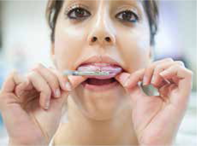
The bite fork is placed with the midline marker coincident with the patient's facial midline and patients are asked to seat the fork firmly using their thumbs (Figure 2).
It is removed and cooled and any interferences on the wax caused by the brackets are carefully trimmed at the chairside using a wax knife.
The bite fork is put back in the mouth to confirm the fit.
Two simple spirit levels are placed on the outer arm of the articulator to record the anteroposterior and transverse planes.
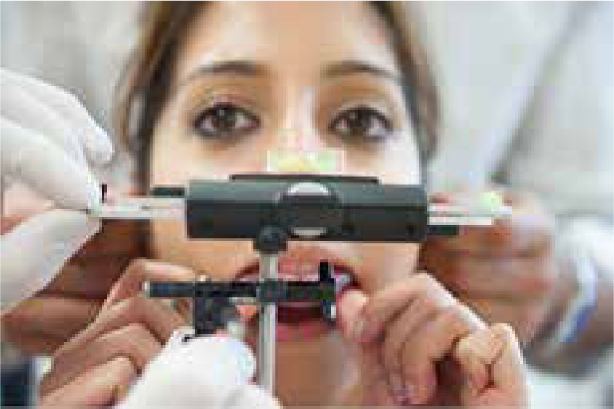
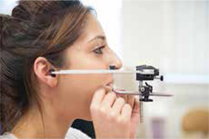
The facebow is positioned and fixed with spirit levels centred (Figures 3 and 4). The levelling of the spirit level in the transverse plane is done by adjusting the seating of one of the ear rods in the external auditory canal.
Figure 1. The use of dental floss to record the patient's facial midline.Figure 2. Wax placed on bite fork seated firmly by the patient in her mouth.Figure 3. Facebow positioned and fixed with bow spirit levels centred.Figure 4. Patient in natural head position and the outer arm of the facebow parallel to the ground.
Once the facebow recording has been taken using this technique the maxillary model can be placed in the articulator at chairside to assess the pitch, roll and yaw of the maxilla. This adds to the diagnosis and can help to inform the patient better of the procedure involved (Figure 5).
Figure 5. Maxillary cast placed on the articulator at chairside to aid diagnosis.
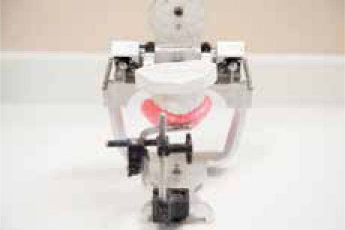
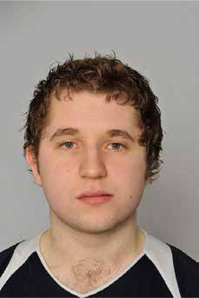
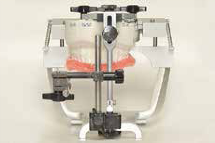
In patients with uneven heights of the external ear, the outer bow is removed from the external ear canal on the ‘abnormal’ side. The transfer jig assembly is then used to transfer the recorded maxillary position onto the articulator in the clinic whilst the patient is still present. The orthodontist and technician can then confirm if any gross maxillary occlusal cant is present. If this stage in the diagnosis confirms a significant undiagnosed cant, the surgeon will be consulted as to whether or not correction of this should be prescribed for the model surgery. Minor lateral shifts of the dental centreline on the articulator can be observed if the ear rods are not equally positioned relative to the patient's mid sagittal plane. Significant lateral shifts can be observed when one ear rod is not placed in the ear at all on one side, as is the case in a patient with a significant panfacial deformity. This is illustrated in an example of a facebow recording taken for a patient with microtia and mild orbital dystopia (Figure 6). The patient's left ear was judged to be abnormally low and had such an atypical anatomy that it would not accept the ear rod. In this situation, the ear rod should be removed from the atypical ear and the facebow recording taken with the spirit level. In such a case, when the casts are mounted the entire model is shifted to the contralateral side of the articulator (Figure 7). This extreme case explains minor lateral shifts in patients with dental centre lines that are coincident with the facial midline. The use of the spirit level to record the transverse occlusal plane allows the accurate assessment of an occlusal cant in the presence of orbital dystopia. In this example, no gross maxillary occlusal cant was present.
Figure 6. Patient with microtia and mild orbital dystopia.Figure 7. Anterior view of significant lateral shift of mounted maxillary model when one ear rod is not seated in external auditory canal.
Summary
Facebow recording and the mounting of maxillary models on articulators can be a source of significant error in patient assessment and treatment planning for orthognathic surgery. In this article the authors have described a simple method of taking a facebow recording with the patient in natural head position using spirit levels. They believe that this technique can help to reduce the pre-surgical error for orthognathic patients and improve clinical results.