Abstract
This case report demonstrates the spontaneous eruption of a maxillary canine from the infra-orbital margin after marsupialization of a dentigerous cyst.
From Volume 6, Issue 2, April 2013 | Pages 58-60
This case report demonstrates the spontaneous eruption of a maxillary canine from the infra-orbital margin after marsupialization of a dentigerous cyst.

Dentigerous cysts are the most common type of developmental odontogenic cysts and make up 20% of all cysts.1 They arise in the follicular tissues covering the fully formed crown of the unerupted tooth. They most commonly involve teeth that are impacted or erupt late. The maxillary permanent canines are the second most commonly affected teeth after the mandibular third molars. Most are detected in adolescents and young adults on routine radiographic examination but there is an increasing prevalence up to the fifth decade. There is a male predilection and the mandible is affected more than the maxilla. This condition commonly presents when a tooth of the permanent series is noted to be missing from the arch. However, by this stage it may have enlarged sufficiently to produce expansion of the jaw. Rarely, when very large, a dentigerous cyst can cause nerve paraesthesia, ophthalmologic signs such as proptosis or epiphora, and nasal symptoms.2 The size can be variable, but a cyst is suspected if the follicular space exceeds 3 mm and can grow to several centimetres in diameter.
Treatment options for dentigerous cysts include enucleation and marsupialization.3 Enucleation is the process of removing the cyst completely without rupture and is generally suitable for small cysts. In large lesions this process might cause tooth devitalization or result in removal of impacted teeth associated with the lesion. Marsupialization consists of making a surgical cavity in the wall of the cyst, which releases the contents, and then maintaining continuity between the cyst and the oral cavity. This allows decompression of the cyst. The ideal treatment for impacted teeth is surgical exposure and orthodontic traction, but spontaneous eruption without orthodontic intervention can occur after extraction of deciduous teeth and cyst marsupialization.4
An 11-year-old boy presented at a specialist orthodontic practice where a large cystic lesion was noted on the initial dental panoramic tomograph (DPT) radiograph. The orthodontist immediately organized an emergency referral to the oral and maxillofacial surgery department.
The patient reported a salty taste in the mouth though he did not have any pain or numbness. He had also noted a lump under his left eye, which had been present for a few months. On examination, the upper left canine and first premolar were unerupted and there was a palpable lump in the upper left quadrant intra-orally.
A biopsy of the cyst was taken under local anaesthesia. The upper left primary canine and first molar were also extracted at this time and an aspirate of cyst fluid taken. The cyst lining was sent for histopathological examination while the cyst fluid was sent for microbiological and biochemical testing. The patient was fitted with a palatal splint to maintain the fistula into the cyst. The histology report was consistent with an inflamed dentigerous cyst.
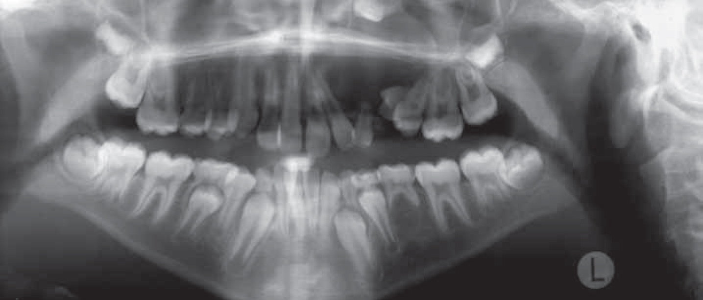
The cyst appeared round and enveloped the upper left canine at its superior margin on the DPT radiograph. The outline was smooth and well defined while the contents were uniformly radiolucent.5 The cyst had displaced the neighbouring teeth, including the upper left lateral incisor, the first and second premolars and the primary canine. There also appeared to be some displacement of the antral floor adjacent to the cyst.
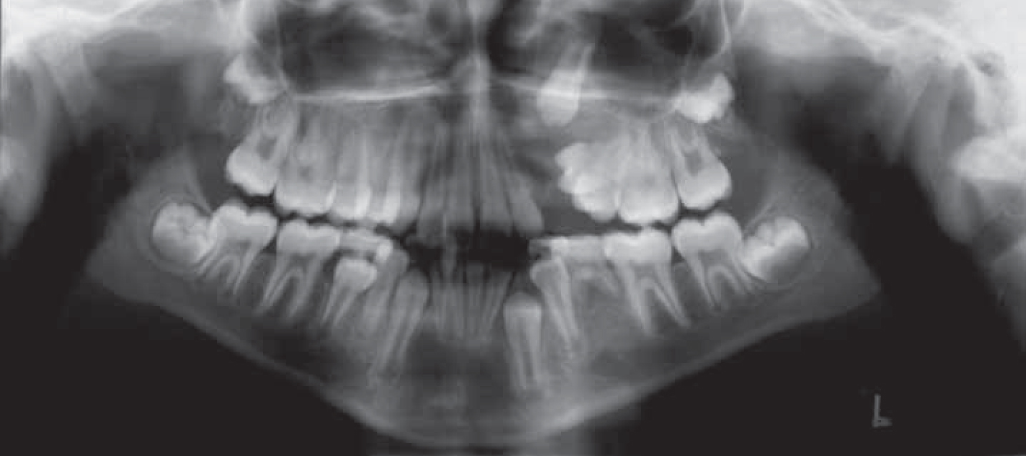
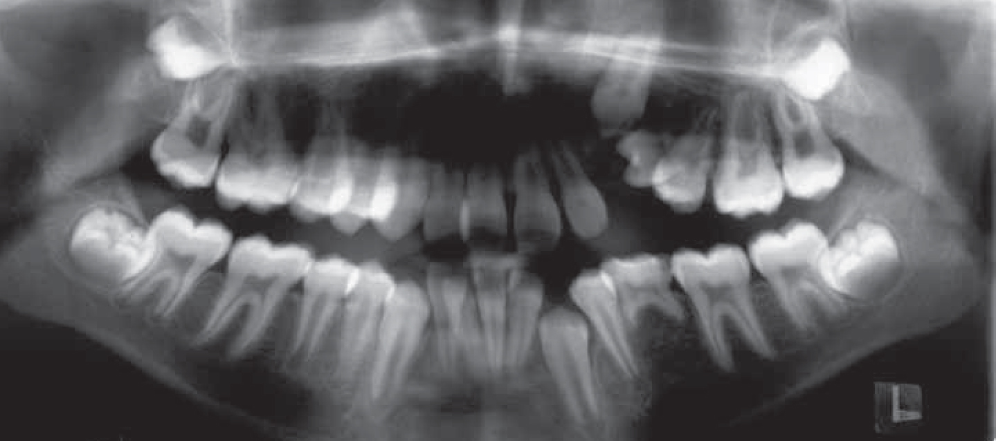
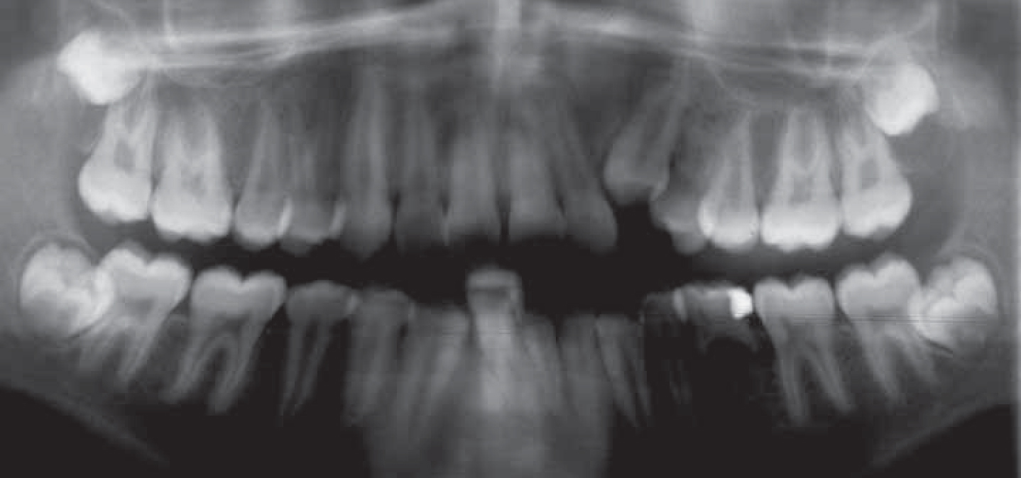
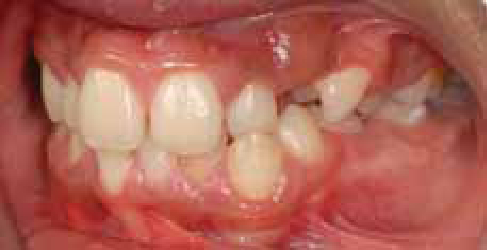
After marsupialization of the cyst, the size of the lesion and canine position were monitored over time with sequential DPTs (Figures 1–4). These revealed that the canine was making steady progress towards the line of the arch. There was rapid progress in the first four months, with the tooth moving half of the distance towards eruption. The radiolucency gradually decreased in size as bony infill occurred. At review three years post marsupialization, the canine was palpable buccally and it was anticipated that it would erupt given adequate space (Figure 5). The upper left first premolar root had a moth-eaten appearance and so it was decided to extract this tooth, along with the lower left second primary molar which had no permanent successor. Second premolars on the right side were also extracted for relief of crowding. Fixed appliances were placed to complete alignment of the upper left canine (Figures 6a, b) and the remainder of the dentition, with eventual debond in March 2011 after 28 months of treatment (Figure 7a, b, c).


Generally, dentigerous cysts are treated by enucleation, however, large cysts that involve multiple teeth can be treated by marsupialization and insertion of a drain. This procedure is less invasive and reduces the risk of damage to the sinuses, nerves and teeth. There are many older reports in the literature of dentigerous cysts in the maxillary sinus being treated by enucleation and removal of the associated impacted teeth. More recently, however, there are successful reports of cases treated by the less invasive marsupialization method, which allows preservation of the displaced teeth.6,7,8 This case demonstrates the great potential for spontaneous relocation of displaced teeth following marsupialization of large dentigerous cysts.